

Effect of Buyang Huanwu Decoction on Delaying Vascular Aging Based on miR-665/DRAM1 Signaling-Mediated Autophagy
-
摘要:
目的 研究补阳还五汤对血管衰老的延缓作用, 探讨其机制是否与microRNA-665(miR-665)/DNA损伤调节自噬调控因子1(DNA damage-regulated autophagy modulator 1, DRAM1)信号介导的自噬有关。 方法 将自然衰老雄性SD大鼠随机分为衰老组, 补阳还五汤低、中、高(9.25、18.5、37.0 g·kg-1)剂量组和白藜芦醇组(80 mg·kg-1), 同时设立年轻组。分离胸主动脉, ELISA法测定血管组织衰老相关β-半乳糖苷酶(Senescence associated β-galactosidase, SA-β-Gal)活性和晚期糖基化终末产物(Advanced glycation end products, AGEs)水平; HE、Masson和EVG染色观察血管组织形态结构; qPCR检测血管组织miR-665表达; 生物信息学分析和双荧光素酶报告基因实验验证miR-665与DRAM1靶向关系; 透射电镜观察血管内自噬小体; Western blot法检测血管组织p16、DRAM1蛋白及自噬相关蛋白LC3、Beclin1和p62的表达; 免疫组织化学法检测血管组织DRAM1的蛋白表达。 结果 与年轻组相比,衰老组大鼠血管中SA-β-Gal活性、AGEs水平和p16蛋白表达增加(P<0.01);血管组织排列紊乱,中膜增厚,胶原纤维增加,弹力纤维出现断裂、紊乱;miR-665基因表达上调(P<0.01);自噬小体数量减少,Beclin1和LC3Ⅱ/Ⅰ蛋白表达降低(P<0.01),p62蛋白表达升高(P<0.01);DRAM1蛋白表达降低(P<0.01)。与衰老组相比,补阳还五汤和白藜芦醇干预能够降低衰老大鼠血管中SA-β-Gal活性(P<0.01)、AGEs水平和p16蛋白表达(P<0.05,P<0.01);改善血管形态和弹力纤维结构,降低血管组织胶原纤维含量。高剂量补阳还五汤明显下调miR-665基因表达(P<0.01),增加血管内自噬小体数量;中剂量和高剂量补阳还五汤明显上调Beclin1和LC3Ⅱ/Ⅰ蛋白表达(P<0.01),下调p62蛋白表达(P<0.05,P<0.01);高剂量补阳还五汤明显上调DRAM1蛋白表达(P<0.05)。生物信息学分析显示,miR-665与DRAM1基因序列存在特异性互补结合位点,双荧光素酶报告实验证实miR-665靶向DRAM1基因并负调控DRAM1蛋白表达。 结论 补阳还五汤可能通过靶向抑制miR-665促进DRAM1蛋白表达, 进而促进血管自噬, 延缓血管衰老。 Abstract:OBJECTIVE This study aimed to investigate the effects of Buyang Huanwu Decoction (BYHWD) on delaying vascular aging and explore whether the underlying mechanism is associated with microRNA-665 (miR-665)/DNA damage-regulated autophagy modulator1 (DRAM1)-mediated autophagy. METHODS Male SD rats with natural aging were randomly divided into the aging group, BYHWD low, medium, high dosage groups (9.25, 18.5, 37.0 g·kg-1) and resveratrol group (80 mg·kg-1), with a young group set as well. The rats in each group were dissected and the blood vessels were collected. ELISA was used to assess senescence associated β-galactosidase (SA-β-Gal) activity and advanced glycation end products (AGEs) level in vascular tissues. HE, Masson, and EVG staining were performed to observe the morphological structure of the vascular tissues. The qPCR was performed to detect the expression of miR-665 in vascular tissues. Bioinformatics analysis and dual-luciferase reporter gene experiments were used to validate the targeting relationship between miR-665 and DRAM1. Transmission electron microscope was used to observe the autophagosome. Western blot was performed to determine the protein expression of p16, DRAM1 and autophagy-related proteins Beclin1, p62 and LC3. Immunohistochemistry was used to detect the protein expression of DRAM1 in vascular tissues. RESULTS Compared to the young group, the aging group showed increased SA-β-Gal activity, AGEs level and p16 protein expression (P < 0.01), disordered arrangement of vascular tissues, thickened media, increased collagen fibers, fractured and disorganized elastic fibers. The expression of miR-665 was upregulated (P < 0.01). The number of autophagosomes was reduced. The protein expression of Beclin1 and LC3Ⅱ/Ⅰ downregulated (P < 0.01), while the protein expression of p62 was upregulated (P < 0.01). In addition, the protein expression of DRAM1 was downregulated in vascular tissues (P < 0.01). Compared to the aging group, intervention with BYHWD and resveratrol reduced SA-β-Gal activity (P < 0.01), AGEs level and p16 protein expression (P < 0.05, P < 0.01), improved vascular morphology and elastic fiber structure, reduced collagen fibers. High dose BYHWD significantly downregulated miR-665 expression (P < 0.01), increased the number of autophagosomes. Medium and high dose of BYHWD significantly upregulated protein expression of Beclin1 and LC3Ⅱ/Ⅰ (P < 0.01), downregulated protein expression of p62 (P < 0.05, P < 0.01). High dose BYHWD significantly upregulated protein expression of DRAM1 in vascular tissues of aging rats (P < 0.05). Bioinformatics analysis revealed the presence of specific complementary binding sites between the sequences of miR-665 and DRAM1. Dual-luciferase reporter assays confirmed that miR-665 targeted DRAM1 gene and negatively regulated DRAM1 protein expression. CONCLUSION BYHWD may promote the protein expression of DRAM1 by inhibiting the expression of miR-665, thereby promoting vascular autophagy and delaying vascular aging. -
Key words:
- Buyang Huanwu Decoction /
- miR-665 /
- DRAM1 /
- autophagy /
- post-transcriptional regulation /
- vascular aging
-

图 1 各组大鼠血管组织SA-β-Gal活性、AGEs水平(n=5)和p16蛋白表达(n=3)
注: 与年轻组比较, * *P<0.01;与衰老组比较, #P<0.05, ##P<0.01。x±s。
Figure 1. SA-β-Gal activity, AGEs levels (n=5) and p16 protein expression (n=3) in vascular tissues of rats in each group

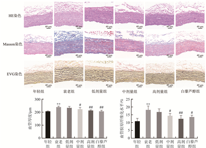
图 2 各组大鼠血管组织HE、Masson和EVG染色
注: 与年轻组比较, * *P<0.01;与衰老组比较, #P<0.05, ##P<0.01。x±s,n=3。
Figure 2. HE, Masson, and EVG staining in vascular tissues of rats in each group

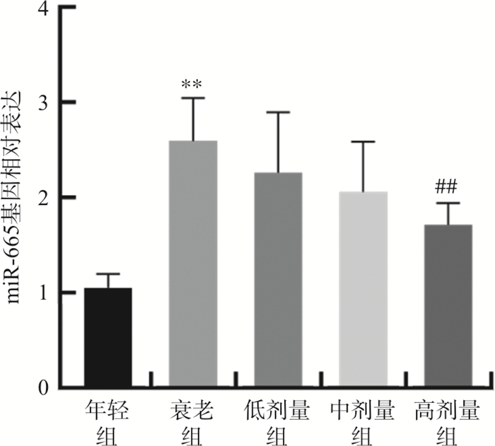
图 3 各组大鼠血管组织miR-665表达
注: 与年轻组比较, * *P<0.01;与衰老组比较, ##P<0.01。x±s,n=5。
Figure 3. Expression of miR-665 in vascular tissues of rats in each group

图 4 各组大鼠血管组织自噬小体数量及LC3、Beclin1、p62蛋白表达
注: 红色箭头代表自噬小体。与年轻组比较, * *P <0.01;与衰老组比较, #P <0.05, ##P <0.01。x±s,n=3。
Figure 4. The number of autophagosomes and the protein expressions of LC3, Beclin1 and p62 in vascular tissues of rats in each group

图 5 miR-665对下游自噬相关蛋白DRAM1的影响
注: 与miR-NC比较, *P<0.05, * *P<0.01。x±s,n=3。
Figure 5. Effect of miR-665 on the downstream autophagy-related protein DRAM1

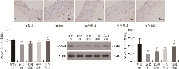
图 6 各组大鼠血管组织DRAM1蛋白表达
注: 与年轻组比较, * *P<0.01;与衰老组比较, #P<0.05。x±s,n=3。
Figure 6. Expression of DRAM1 protein in vascular tissues of rats in each group

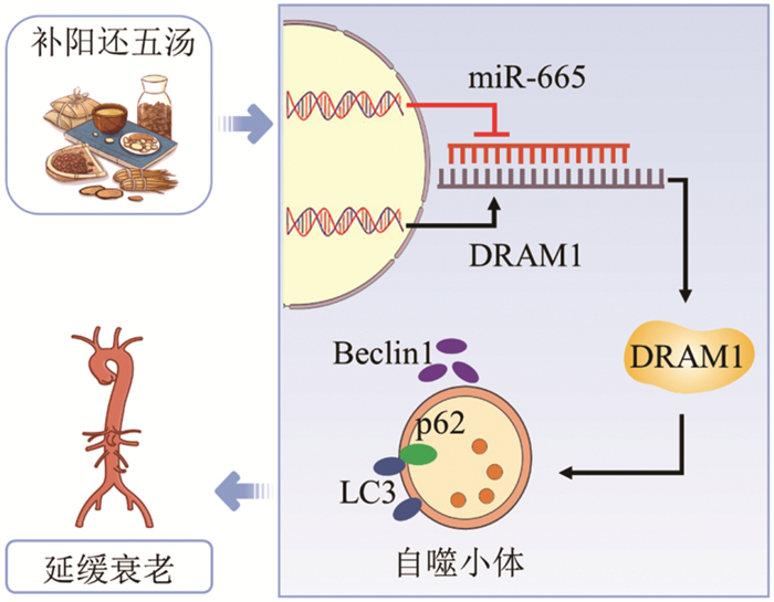
图 7 补阳还五汤延缓血管衰老的可能机制
Figure 7. Possible mechanism of Buyang Huanwu Decoction delaying vascular aging
表 1 引物序列
Table 1. Primer sequences
基因 序列(5'→3') miR-665 F: ACACTCCAGCTGGGACCAGGAGGCUGAGGUCC
R: CTCAACTGGTGTCGTGGAGTCGGCAATTCAGTTGAGTAAGGGACU6 F: CTCGCTTCGGCAGCACA
R: AACGCTTCACGAATTTGCGTAll R: TCAACTGGTGTCGTGGA  下载: 导出CSV
-
[1] LAROCCA T J, MARTENS C R, SEALS D R. Nutrition and other lifestyle influences on arterial aging[J]. Ageing Res Rev, 2017, 39: 106-119. doi: 10.1016/j.arr.2016.09.002 [2] DONATO A J, MACHIN D R, LESNIEWSKI L A. Mechanisms of dysfunction in the aging vasculature and role in age-related disease[J]. Circ Res, 2018, 123(7): 825-848. doi: 10.1161/CIRCRESAHA.118.312563 [3] WANG M Y, ZHANG L, ZHU W Q, et al. Calorie restriction curbs proinflammation that accompanies arterial aging, preserving a youthful phenotype[J]. J Am Heart Assoc, 2018, 7(18): e009112. doi: 10.1161/JAHA.118.009112 [4] 颜德馨, 胡泉林, 王平平, 等. 气虚血瘀是人体衰老的主要机制[J]. 中国医药学报, 1989, 4(2): 10-12, 79. doi: 10.3321/j.issn:1673-1727.1989.02.002YAN D X, HU Q L, WANG P P, et al. Deficiency of qi and stagnancy of blood—An important mechanism of PhysicaI senility[J]. China J Tradit Chin Med Pharm, 1989, 4(2): 10-12, 79. doi: 10.3321/j.issn:1673-1727.1989.02.002 [5] 戚璐, 谢长. 益气活血法延缓衰老理论探源[J]. 湖北中医药大学学报, 2012, 14(1): 46-47. doi: 10.3969/j.issn.1008-987x.2012.01.19QI L, XIE C. Probe into the theory of delaying aging by supplementing qi and activating blood circulation[J]. J Hubei Univ Chin Med, 2012, 14(1): 46-47. doi: 10.3969/j.issn.1008-987x.2012.01.19 [6] TAO L L, LEI Y, WANG G L, et al. Effect of extracts from Radix Ginseng, Radix Notoginseng and Rhizoma Chuanxiong on delaying aging of vascular smooth muscle cells in aged rats[J]. Chin J Integr Med, 2012, 18(8): 582-590. doi: 10.1007/s11655-012-1180-1 [7] 张伟, 贺冰, 李亮, 等. 补阳还五汤促进内皮祖细胞修复损伤血管内皮[J]. 中国病理生理杂志, 2017, 33(11): 1969-1974. https://www.cnki.com.cn/Article/CJFDTOTAL-ZBLS201711010.htmZHANG W, HE B, LI L, et al. BYHWD promotes endothelial progenitor cells to repair damaged vascular endothelium[J]. Chin J Pathophysiol, 2017, 33(11): 1969-1974. https://www.cnki.com.cn/Article/CJFDTOTAL-ZBLS201711010.htm [8] 洪允祥, 鲍军, 楼建国, 等. 补阳还五汤延缓衰老的临床研究[J]. 中国中西医结合杂志, 1994, 14(增1): 87-89, 427. https://www.cnki.com.cn/Article/CJFDTOTAL-ZZXJ1994S1041.htmHONG Y X, BAO J, LOU J G, et al. Clinical study on delaying aging with Buyanghuanwu Decoction[J]. Chin J Integr Tradit West Med, 1994, 14(Suppl 1): 87-89, 427. https://www.cnki.com.cn/Article/CJFDTOTAL-ZZXJ1994S1041.htm [9] 华润龄, 任光荣, 王明武, 等. 益气活血液抗衰老作用的临床研究[J]. 南京中医药大学学报, 1996, 12(5): 16-18. https://www.cnki.com.cn/Article/CJFDTOTAL-NJZY605.007.htmHUA R L, REN G R, WANG M W, et al. Anti-ageing effects of qi-supplementing and blood-activating liquid[J]. J Nanjing Univ Tradit Chin Med, 1996, 12(5): 16-18. https://www.cnki.com.cn/Article/CJFDTOTAL-NJZY605.007.htm [10] 孙正骥. 补阳还五汤苷类组分抗内皮祖细胞衰老的作用机制[D]. 长沙: 湖南中医药大学, 2021.SUN Z J. Anti-aging Mechanism of Glycosides from Buyanghuanwu Decoction on Endothelial Progenitor Cells[D]. Changsha: Hunan University of Chinese Medicine, 2021. [11] ZHANG L, WEI C S, RUAN Y J, et al. Serum containing Buyang Huanwu Decoction prevents age-associated migration and invasion of human vascular smooth muscle cells by up regulating SIRT1 expression[J]. Biosci Trends, 2018, 12(3): 282-290. doi: 10.5582/bst.2018.01063 [12] 张雅楠, 高劲松, 张莉. 补阳还五汤延缓血管平滑肌细胞衰老的机制研究[J]. 世界科学技术-中医药现代化, 2020, 22(10): 3658-3664. https://www.cnki.com.cn/Article/CJFDTOTAL-SJKX202010035.htmZHANG Y N, GAO J S, ZHANG L. The mechanism of Buyang Huanwu Decoction in preventing the aging of vascular smooth muscle cells[J]. Mod Tradit Chin Med Mater Med World Sci Technol, 2020, 22(10): 3658-3664. https://www.cnki.com.cn/Article/CJFDTOTAL-SJKX202010035.htm [13] XU F, ZHONG J Y, LIN X, et al. Melatonin alleviates vascular calcification and ageing through exosomal miR-204/miR-211 cluster in a paracrine manner[J]. J Pineal Res, 2020, 68(3): e12631. doi: 10.1111/jpi.12631 [14] TAI S, HU X Q, PENG D Q, et al. The roles of autophagy in vascular smooth muscle cells[J]. Int J Cardiol, 2016, 211: 1-6. doi: 10.1016/j.ijcard.2016.02.128 [15] JIANG F. Autophagy in vascular endothelial cells[J]. Clin Exp Pharmacol Physiol, 2016, 43(11): 1021-1028. doi: 10.1111/1440-1681.12649 [16] ZHANG Y, LIANG Q Y, ZHANG Y N, et al. Olmesartan alleviates bleomycin-mediated vascular smooth muscle cell senescence via the miR-665/SDC1 axis[J]. Am J Transl Res, 2020, 12(9): 5205-5220. [17] CHEN T B, LIANG Q Y, XU J L, et al. MiR-665 regulates vascular smooth muscle cell senescence by interacting with LncRNA GAS5/SDC1[J]. Front Cell Dev Biol, 2021, 9: 700006. doi: 10.3389/fcell.2021.700006 [18] 祝晓玲, 刘淑梅, 杨爱华, 等. MiR-665在宫腔粘连及宫颈癌患者中的表达及意义[J]. 广东医学, 2020, 41(18): 1851-1857. https://www.cnki.com.cn/Article/CJFDTOTAL-GAYX202018005.htmZHU X L, LIU S M, YANG A H, et al. The expression and clinical significance of miR-665 in intrauterine adhesion and cervical cancer[J]. Guangdong Med J, 2020, 41(18): 1851-1857. https://www.cnki.com.cn/Article/CJFDTOTAL-GAYX202018005.htm [19] CRIGHTON D, WILKINSON S, O'PREY J, et al. DRAM, a p53-induced modulator of autophagy, is critical for apoptosis[J]. Cell, 2006, 126(1): 121-134. doi: 10.1016/j.cell.2006.05.034 [20] ZHU C L, ZHANG L, ZHENG Y, et al. Effects of estrogen on stress-induced premature senescence of vascular smooth muscle cells: A novel mechanism for the "time window theory" of menopausal hormone therapy[J]. Atherosclerosis, 2011, 215(2): 294-300. doi: 10.1016/j.atherosclerosis.2010.12.025 [21] 严翼, 张浩, 杨志健, 等. 雌激素缺乏对心肌衰老、凋亡和心功能的影响[J]. 医学研究杂志, 2018, 47(12): 110-114. https://www.cnki.com.cn/Article/CJFDTOTAL-YXYZ201812028.htmYAN Y, ZHANG H, YANG Z J, et al. Research of estrogen deficiency on myocardial aging, apoptosis and cardiac function[J]. J Med Res, 2018, 47(12): 110-114. https://www.cnki.com.cn/Article/CJFDTOTAL-YXYZ201812028.htm [22] MOHAMAD KAMAL N S, SAFUAN S, SHAMSUDDIN S, et al. Aging of the cells: Insight into cellular senescence and detection Methods[J]. Eur J Cell Biol, 2020, 99(6): 151108. doi: 10.1016/j.ejcb.2020.151108 [23] 孙红艳, 刘洪臣. 晚期糖基化终末产物(AGEs)与衰老[J]. 中华老年口腔医学杂志, 2010, 8(5): 314-317. doi: 10.3969/j.issn.1672-2973.2010.05.019SUN H Y, LIU H C. The correlation of the Advanced glycosylation and products and aging[J]. Chin J Geriatr Dent, 2010, 8(5): 314-317. doi: 10.3969/j.issn.1672-2973.2010.05.019 [24] LIU J Y, SOUROULLAS G P, DIEKMAN B O, et al. Cells exhibiting strong p16INK4a promoter activation in vivo display features of senescence[J]. Proc Natl Acad Sci USA, 2019, 116(7): 2603-2611. doi: 10.1073/pnas.1818313116 [25] LAPIERRE L R, KUMSTA C, SANDRI M, et al. Transcriptional and epigenetic regulation of autophagy in aging[J]. Autophagy, 2015, 11(6): 867-880. doi: 10.1080/15548627.2015.1034410 [26] 张茹鑫, 李承罡, 杜若琛, 等. 人脐带间充质干细胞对自然衰老大鼠海马自噬水平的影响[J]. 中国实验动物学报, 2020, 28(6): 796-804. https://www.cnki.com.cn/Article/CJFDTOTAL-ZGSD202006009.htmZHANG R X, LI C G, DU R C, et al. Effects of human umbilical cord mesenchymal stem cells on autophagy in the naturally aging rat hippocampus[J]. Acta Lab Animalis Sci Sin, 2020, 28(6): 796-804. https://www.cnki.com.cn/Article/CJFDTOTAL-ZGSD202006009.htm [27] HYTTINEN J M T, BLASIAK J, FELSZEGHY S, et al. MicroRNAs in the regulation of autophagy and their possible use in age-related macular degeneration therapy[J]. Ageing Res Rev, 2021, 67: 101260. doi: 10.1016/j.arr.2021.101260 [28] 冀小伟, 张连城. 中医对衰老的认识[J]. 中医杂志, 2013, 54(17): 1527-1529. https://www.cnki.com.cn/Article/CJFDTOTAL-ZZYZ201317042.htmJI X W, ZHANG L C. Understanding of aging in traditional Chinese medicine[J]. J Tradit Chin Med, 2013, 54(17): 1527-1529. https://www.cnki.com.cn/Article/CJFDTOTAL-ZZYZ201317042.htm [29] 邸睿宁, 姜欢欢, 李亚茹, 等. 黄芪-当归配伍对D-半乳糖致衰老小鼠抗氧化的影响[J]. 陕西中医药大学学报, 2023, 46(2): 100-104. https://www.cnki.com.cn/Article/CJFDTOTAL-SXXY202302016.htmDI R N, JIANG H H, LI Y R, et al. Effect of astragalus-angelica compatibility on antioxidation in aging mice induced by D-galactose[J]. J Shaanxi Univ Chin Med, 2023, 46(2): 100-104. https://www.cnki.com.cn/Article/CJFDTOTAL-SXXY202302016.htm [30] MIN F, SUN H Q, WANG B, et al. Hepatoprotective effects of hydroxysafflor yellow A in D-galactose-treated aging mice[J]. Eur J Pharmacol, 2020, 881: 173214. doi: 10.1016/j.ejphar.2020.173214 [31] 刘颖, 李花, 刘旺华, 等. 川芎对D-半乳糖衰老模型小鼠的抗衰老作用研究[J]. 湖南中医杂志, 2021, 37(2): 147-149. https://www.cnki.com.cn/Article/CJFDTOTAL-HNZO202102053.htmLIU Y, LI H, LIU W H, et al. Anti-senescence mechanism of Rhizoma Ligustici Chuanxiong in a mouse model of senescence induced by D-galactose[J]. Hunan J Tradit Chin Med, 2021, 37(2): 147-149. https://www.cnki.com.cn/Article/CJFDTOTAL-HNZO202102053.htm [32] FAN J H, LI H P, NIE X, et al. MiR-665 aggravates heart failure via suppressing CD34-mediated coronary microvessel angiogenesis[J]. Aging, 2018, 10(9): 2459-2479. doi: 10.18632/aging.101562 [33] LIU C Z, TANG M M, ZHANG X Q, et al. Knockdown of miR-665 protects against cardiomyocyte ischemia/reperfusion injury-induced ROS accumulation and apoptosis through the activation of Pak1/akt signaling in myocardial infarction[J]. Int Heart J, 2020, 61(2): 347-354. doi: 10.1536/ihj.19-416 [34] LI W, HE P C, HUANG Y G, et al. Selective autophagy of intracellular organelles: Recent research advances[J]. Theranostics, 2021, 11(1): 222-256. doi: 10.7150/thno.49860 [35] YUAN J, ZHANG Q Y, CHEN S H, et al. LC3-associated phagocytosis in bacterial infection[J]. Pathogens, 2022, 11(8): 863. doi: 10.3390/pathogens11080863 [36] JEONG S J, ZHANG X Y, RODRIGUEZ-VELEZ A, et al. p62/ SQSTM1 and selective autophagy in cardiometabolic diseases[J]. Antioxid Redox Signal, 2019, 31(6): 458-471. doi: 10.1089/ars.2018.7649 [37] KANG C, XU Q K, MARTIN T D, et al. The DNA damage response induces inflammation and senescence by inhibiting autophagy of GATA4[J]. Science, 2015, 349(6255): 5612. [38] LI W W, WANG H J, TAN Y Z, et al. Reducing lipofuscin accumulation and cardiomyocytic senescence of aging heart by enhancing autophagy[J]. Exp Cell Res, 2021, 403(1): 112585. [39] CARNIO S, LOVERSO F, BARAIBAR M A, et al. Autophagy impairment in muscle induces neuromuscular junction degeneration and precocious aging[J]. Cell Rep, 2014, 8(5): 1509-1521. [40] LI H, PENG D, ZHANG S J, et al. Buyang Huanwu Decoction promotes neurogenesis via sirtuin 1/autophagy pathway in a cerebral ischemia model[J]. Mol Med Rep, 2021, 24(5): 791. [41] 张扬, 严寒, 梁永. 补阳还五汤对缺血性脑卒中大鼠认知功能的影响[J]. 中成药, 2023, 45(4): 1309-1314. https://www.cnki.com.cn/Article/CJFDTOTAL-ZCYA202304048.htmZHANG Y, YAN H, LIANG Y. Effect of buyanghuanwu decoction on cognitive function in rats with ischemic stroke[J]. Chin Tradit Pat Med, 2023, 45(4): 1309-1314. https://www.cnki.com.cn/Article/CJFDTOTAL-ZCYA202304048.htm [42] YANG J R, CHEN D P, HE Y N, et al. MiR-34 modulates Caenorhabditis elegans lifespan via repressing the autophagy gene atg9[J]. Age (Dordr), 2013, 35(1): 11-22. [43] LIU X J, FU B, CHEN D P, et al. MiR-184 and miR-150 promote renal glomerular mesangial cell aging by targeting Rab1a and Rab31[J]. Exp Cell Res, 2015, 336(2): 192-203. [44] GUO Q Q, LIN Y, HU J Z. Inhibition of miR-665-3p enhances autophagy and alleviates inflammation in Fusarium solani -induced keratitis[J]. Invest Ophthalmol Vis Sci, 2021, 62(1): 24. [45] LI Z L, WANG G Z, FENG D C, et al. Targeting the miR-665-3p-ATG4B-autophagy axis relieves inflammation and apoptosis in intestinal ischemia/reperfusion[J]. Cell Death Dis, 2018, 9(5): 483. [46] YU M Q, JIANG Y G, FENG Q L, et al. DRAM1 protects neuroblastoma cells from oxygen-glucose deprivation/reperfusion-induced injury via autophagy[J]. Int J Mol Sci, 2014, 15(10): 19253-19264. [47] HU W L, CHEN S, THORNE R F, et al. TP53, TP53 target genes (DRAM, TIGAR), and autophagy[J]. Adv Exp Med Biol, 2019, 1206: 127-149. -


 点击查看大图

计量
- 文章访问数: 38
- HTML全文浏览量: 1
- PDF下载量: 8
- 被引次数: 0
