

Research on the Protective Effects and Mechanisms of Cycloartenol, the Effective Component of Pinellia Ternata, on Myocardial Ischemia-Reperfusion Injury in Mice
-
摘要:
目的 探究半夏有效成分环阿屯醇对小鼠心肌缺血再灌注损伤(Myocardial ischemia reperfusion injury, MIRI)的影响及作用机制。 方法 在体外实验中, 从1~3日龄的SD雄性大鼠乳鼠上提取原代心肌细胞, 分为对照(Control)组、缺氧/复氧(Hypoxia/Reoxygenation,H/R)组、环阿屯醇低剂量组(3 μmol·L-1)、环阿屯醇高剂量组(10 μmol·L-1)和SB203580组。各组预给药干预后,在缺氧培养箱中缺氧培养3 h, 正常培养箱中复氧培养3 h复制缺氧/复氧模型。利用CCK-8检测各组心肌细胞活力, 流式法检测细胞凋亡率, Western blot检测p38 MAPK、p-p38 MAPK的表达水平。在体内实验中, 将7周龄的雄性C57BL/6J小鼠随机分为对照(Control)组, 缺血/再灌注损伤(Ischemia/Reperfusion, I/R)组, 环阿屯醇低、中、高(0.2、0.5、1.0 mg·kg-1)剂量组。手术前7 d连续给药, 通过小鼠心脏冠状动脉左前降支(Left anterior descending artery, LAD)结扎30 min, 再灌注24 h的方式制备MIRI模型。小动物超声心动图检测左心室射血分数(Left ventricular ejection fraction, LVEF)、小鼠心输出量(Cardiac output, CO)、左室缩短分数(Left ventricular fractional shortening, LVFS); TTC染色法检测各组小鼠心脏的缺血梗死面积的变化; HE染色法观察各组小鼠心肌组织的变化; Western blot检测小鼠心肌组织中p38 MAPK、p-p38 MAPK、IL-6及TNF-α的表达水平; ELISA法检测小鼠血清肌酸激酶同工酶(CK-MB)、乳酸脱氢酶(LDH)、肌钙蛋白I(cTnI)、细胞白细胞介素-6(IL-6)、肿瘤坏死因子-α(TNF-α)水平。 结果 体外实验中, 与H/R组相比, 环阿屯醇预处理组和SB203580组的心肌细胞活力有明显提高(P < 0.05), 细胞凋亡率有所下降(P < 0.05), 可以下调IL-6表达水平(P < 0.01), 降低p-p38 MAPK/p38 MAPK的比值(P < 0.05, P < 0.01)。体内实验证明, 与I/R组相比, 中、高剂量环阿屯醇预处理可以显著提高LVEF、LVFS及CO值(P < 0.05, P < 0.01), 减少心肌缺血梗死面积(P < 0.05), 提高心肌功能, 有效保护心肌组织纤维, 下调小鼠血清中CK-MB、LDH、cTnI、IL-6和TNF-α水平(P < 0.05);环阿屯醇高剂量组降低p-p38 MAPK/p38 MAPK的比值(P < 0.05), 减少IL-6和TNF-α的表达水平(P < 0.05)。 结论 环阿屯醇预处理可以保护小鼠心功能, 减轻MIRI, 其作用机制可能与抑制p38 MAPK磷酸化, 减轻炎性反应有关。 Abstract:OBJECTIVE To explore the effects and mechanisms of cycloartenol on myocardial ischemia-reperfusion injury in mice. METHODS In vitro experiments, primary cardiomyocytes were extracted from 1-3 days SD mice. The hypoxia/reoxygenation model was established by incubating cells in a hypoxic culture box for 3 hours followed by reoxygenation in a normal culture box for 3 hours. The primary cardiomyocytes were divided into Control group, H/R group, low-dose (3 μmol·L-1) and high-dose (10 μmol·L-1) cycloartenol groups, and SB203580 group. CCK-8 was used to detect cell viability, the apoptosis rate was detected by flow cytometry, and Western blot was used to detect the expression levels of p38 MAPK and p-p38 MAPK in each group. In an in vivo experiment, 7-week-old male C57BL/6J mice were randomly divided into a Control group, an I/R group, and three doses of cycloartenol (0.2, 0.5, 1.0 mg·kg-1) groups. The mice were continuously administered for seven days before the surgery. The model was prepared by ligation of the left anterior descending coronary artery (LAD) for 30 minutes, followed by reperfusion for 24 hours to induce myocardial ischemia-reperfusion injury. Left ventricular ejection fraction (LVEF), cardiac output (CO), left ventricular fractional shortening (LVFS) of each group of mice were detected by small animal ultrasound. TTC staining was used to detect the changes of ischemic infarct size in each group. The changes of myocardial tissue in each group were observed by HE staining. The expression levels of p38 MAPK, p-p38 MAPK, IL-6 and TNF-α in myocardial tissue of mice were detected by Western blot. Serum levels of creatine kinase isoenzyme (CK-MB), lactate dehydrogenase (LDH), cardiac troponin I (cTnI), interleukin-6 (IL-6), and tumor necrosis factor-α (TNF-α) were measured using ELISA kits. RESULTS In vitro experiments demonstrated that compared with the H/R group, both the cycloartenol and SB203580 pretreatment groups showed a significant increase in myocardial cell viability and the apoptosis rate decrease, which can downregulate the protein expression level of p-p38 MAPK and decrease the ratio of p-p38 MAPK/p38 MAPK(P < 0.05). In vivo experiments confirmed that compared with the I/R group, cycloartenol pretreatment significantly improved LVEF, LVFS, and CO values (P < 0.05), reduce myocardial ischemic infarct size, thereby enhancing myocardial function. The protein expression level of p-p38 MAPK in myocardial tissue was down-regulated, the ratio of p-p38 MAPK/p38 MAPK was decreased, and the expression levels of IL-6 and TNF-α were decreased. Additionally, cycloartenol pretreatment reduced the levels of CK-MB, LDH, cTnI, IL-6, and TNF-α in mouse serum (P < 0.05). CONCLUSION Pre-treatment with cycloartenol can protect mouse cardiac function and alleviate myocardial ischemia-reperfusion injury. Its mechanism of action may be related to the inhibition of p38 MAPK phosphorylation, reducing inflammatory reactions. -
Key words:
- cycloartenol /
- hypoxia/reoxygenation /
- ischemia/reperfusion injury /
- p38 MAPK /
- inflammatory response
-

图 1 不同浓度环阿屯醇预处理对H/R心肌细胞活力的影响
注: 与0 μmol·L-1组比较, *P < 0.05, **P < 0.01。x±s,n=3。
Figure 1. Effect of different concentrations of cycloartenol pretreatment on H/R cardiomyocyte viability

图 2 不同药物预处理对各组心肌细胞活力的影响
注: 与Control组相比, *P < 0.05;与H/R组相比, #P < 0.05。x±s,n=4。
Figure 2. Effects of different drug preconditioning on myocardial cell viability in each group

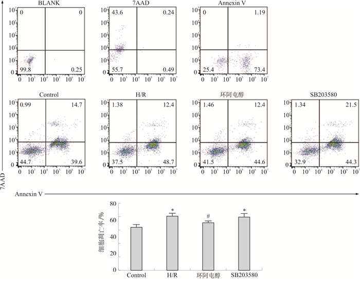
图 3 环阿屯醇预处理对各组心肌细胞凋亡率的影响
注: 与Control组相比, *P < 0.05;与H/R组相比, #P < 0.05。x±s,n=3。
Figure 3. Effect of cycloartenol pretreatment on apoptosis of cardiomyocytes in each group

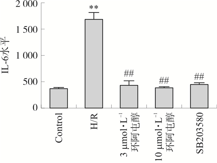
图 4 各组IL-6水平的比较
注: 与Control组相比, **P < 0.01;与H/R组相比, ##P < 0.01。x±s,n=4。
Figure 4. IL-6 levels in each group

图 5 各组p-p38 MAPK/p38 MAPK表达量的比较
注: 与Control组相比, *P < 0.05, 与H/R组相比, #P < 0.05,##P < 0.01。x±s,n=3。
Figure 5. The change of p-p38 MAPK/p38 MAPK in each group

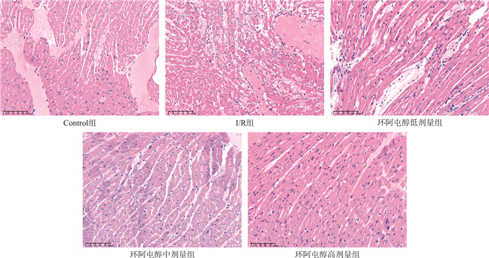
图 7 各组小鼠HE染色示意图(×800)
Figure 7. Schematic diagram of HE staining in each group of mice (×800)

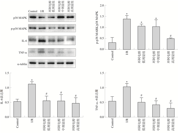
图 8 各组小鼠心肌组织p-p38 MAPK/p38 MAPK、IL-6、TNF-α蛋白表达的比较
注: 与Control组相比, *P < 0.05, 与I/R组相比, #P < 0.05。x±s,n=4。
Figure 8. Comparison of p-p38 MAPK/p38 MAPK, IL-6, TNF-α protein expression in myocardial tissue of mice in each group
表 1 环阿屯醇预处理对小鼠LVFS、LVEF及CO的影响(x±s,n=10)
Table 1. Effects of cycloartenol pretreatment on LVFS, LVEF and CO in mice (x±s, n=10)
组别 剂量/(mg·kg-1) LVFS/% LVEF/% CO/(mL·min-1) Control组 - 34.25±3.85 64.39±5.59 25.71±5.52 I/R组 - 15.59±2.67* 33.80±5.34* 15.47±2.20* 环阿屯醇低剂量组 0.2 19.03±4.45* 39.80±8.08* 21.69±1.21# 环阿屯醇中剂量组 0.5 25.53±4.73*# 51.29±8.13*# 21.71±1.05# 环阿屯醇高剂量组 1.0 30.82±5.92## 59.41±8.78## 21.96±2.67# 注: 与Control组比较, *P < 0.05;与I/R组比较, #P < 0.05, ##P < 0.01。  下载: 导出CSV
表 2 各组IA/HA统计结果(x±s, n=4)
Table 2. Statistical result of IA/HA in each group(x±s, n=4)
组别 剂量/(mg·kg-1) IA/HA Control组 - 0.04±0.04 I/R组 - 0.34±0.03* 环阿屯醇低剂量组 0.2 0.27±0.01* 环阿屯醇中剂量组 0.5 0.19±0.01#* 环阿屯醇高剂量组 1.0 0.15±0.01#* 注: 与Control组比较, *P < 0.05;与I/R组比较, #P < 0.05。
下载: 导出CSV
表 3 环阿屯醇预处理对小鼠血清CK-MB、LDH水平的影响(x±s,ng·mL-1,n=5)
Table 3. Effects of cycloartenol pretreatment on serum CK-MB and LDH of mice (x±s, ng·mL-1, n=5)
组别 剂量/(mg·kg-1) CK-MB LDH Control组 - 67.84±6.09 7.36±0.18 I/R组 - 240.38±27.04* 10.74±0.54* 环阿屯醇低剂量组 0.2 181.57±9.59*# 9.53±0.18*# 环阿屯醇中剂量组 0.5 145.07±8.82*# 8.95±0.24*# 环阿屯醇高剂量组 1.0 103.63±6.61*# 8.04±0.26*# 注: 与Control组比较, *P < 0.05;与I/R组比较, #P < 0.05。
下载: 导出CSV
表 4 环阿屯醇预处理对小鼠血清IL-6、TNF-α和cTnI水平的影响(x±s,n=5)
Table 4. Effects of cycloartenol pretreatment on serum IL-6, TNF-α and cTnI in mice (x±s, n=5)
组别 剂量/(mg·kg-1) IL-6/(pg·mL-1) TNF-α/(pg·mL-1) cTnI/(ng·mL-1) Control组 - 465.48±22.12 167.15±21.98 3.46±0.15 I/R组 - 1 150.80±300.31* 322.14±24.54* 9.68±1.30* 环阿屯醇低剂量组 0.2 872.11±84.23*# 293.53±6.10*# 7.31±1.00*# 环阿屯醇中剂量组 0.5 719.00±26.60*# 249.78±10.82*# 5.7±0.29*# 环阿屯醇高剂量组 1.0 532.37±14.70# 216.8±29.60*# 4.25±0.35# 注: 与Control组比较, *P < 0.05;与I/R组比较, #P < 0.05。
下载: 导出CSV
-
[1] ZHAO Z Q. Postconditioning in reperfusion injury: A status report[J]. Cardiovasc Drugs Ther, 2010, 24(3): 265-279. doi: 10.1007/s10557-010-6240-1 [2] MOENS A L, CLAEYS M J, TIMMERMANS J P, et al. Myocardial ischemia/reperfusion-injury, a clinical view on a complex pathophysiological process[J]. Int J Cardiol, 2005, 100(2): 179-190. doi: 10.1016/j.ijcard.2004.04.013 [3] 韩军花. 植物甾醇的性质、功能及应用[J]. 国外医学(卫生学分册), 2001(5): 285-291.HAN J H. Properties, functions and applications of phytosterols[J]. Foreign Med Sci Sect Hyg, 2001(5): 285-291. [4] DE JONG A, PLAT J, MENSINK R P. Metabolic effects of plant sterols and stanols[J]. J Nutr Biochem, 2003, 14(7): 362-369. doi: 10.1016/S0955-2863(03)00002-0 [5] BLANCO-SALAS J, VAZQUEZ F M, HORTIGON-VINAGRE M P, et al. Bioactive phytochemicals from Mercurialis spp. used in traditional Spanish medicine[J]. Plants, 2019, 8(7): 193. doi: 10.3390/plants8070193 [6] EZZAT S M, CHOUCRY M A, KANDIL Z A. Antibacterial, antioxidant, and topical anti-inflammatory activities of Bergia ammannioides : A wound-healing plant[J]. Pharm Biol, 2016, 54(2): 215-224. doi: 10.3109/13880209.2015.1028079 [7] YANG K L, ZENG L T, GE A Q, et al. Exploring the oxidative stress mechanism of Buyang Huanwu Decoction in intervention of vascular dementia based on systems biology strategy[J]. Oxid Med Cell Longev, 2021, 2021: 8879060. [8] AWAD A B, DOWNIE A, FINK C S, et al. Dietary phytosterol inhibits the growth and metastasis of MDA-MB-231 human breast cancer cells grown in SCID mice[J]. Anticancer Res, 2000, 20(2A): 821-824. [9] AWAD A B, DOWNIE A C, FINK C S. Inhibition of growth and stimulation of apoptosis by beta-sitosterol treatment of MDA-MB-231 human breast cancer cells in culture[J]. Int J Mol Med, 2000, 5(5): 541-545. [10] JANEZIC S A, RAO A V. Dose-dependent effects of dietary phytosterol on epithelial cell proliferation of the murine colon[J]. Food Chem Toxicol, 1992, 30(7): 611-616. doi: 10.1016/0278-6915(92)90195-Q [11] DE STEFANI E, BOFFETTA P, RONCO A L, et al. Plant sterols and risk of stomach cancer: A case-control study in Uruguay[J]. Nutr Cancer, 2000, 37(2): 140-144. doi: 10.1207/S15327914NC372_4 [12] NIU H F, LI X M, YANG A J, et al. Cycloartenol exerts anti-proliferative effects on Glioma U87 cells via induction of cell cycle arrest and p38 MAPK-mediated apoptosis[J]. J BUON, 2018, 23(6): 1840-1845. [13] ISLAM M S, YOSHIDA H, MATSUKI N, et al. Antioxidant, free radical-scavenging, and NF-κB-inhibitory activities of phytosteryl ferulates: Structure-activity studies[J]. J Pharmacol Sci, 2009, 111(4): 328-337. doi: 10.1254/jphs.09146FP [14] SULTANA S, ALAM A, KHAN N, et al. Inhibition of benzoyl peroxide and ultraviolet-B radiation induced oxidative stress and tumor promotion markers by cycloartenol in murine skin[J]. Redox Rep, 2003, 8(2): 105-112. doi: 10.1179/135100003125001422 [15] BADREDDINE A, ZARROUK A, KARYM E M, et al. Argan oil-mediated attenuation of organelle dysfunction, oxidative stress and cell death induced by 7-ketocholesterol in murine oligodendrocytes 158N[J]. Int J Mol Sci, 2017, 18(10): 2220. doi: 10.3390/ijms18102220 [16] AMALRAJ S, KRUPA J, SRIRAMAVARATHARAJAN V, et al. Chemical characterization, antioxidant, antibacterial and enzyme inhibitory properties of Canthium coromandelicum, a valuable source for bioactive compounds[J]. J Pharm Biomed Anal, 2021, 192: 113620. doi: 10.1016/j.jpba.2020.113620 [17] JUNG H A, JUNG Y J, HYUN S K, et al. Selective cholinesterase inhibitory activities of a new monoterpene diglycoside and other constituents from Nelumbo nucifera stamens[J]. Biol Pharm Bull, 2010, 33(2): 267-272. doi: 10.1248/bpb.33.267 [18] NAIR A N S, NAIR R V R, NAIR A P R, et al. Antidiabetes constituents, cycloartenol and 24-methylenecycloartanol, from Ficus krishnae[J]. PLoS ONE, 2020, 15(6): e0235221. doi: 10.1371/journal.pone.0235221 [19] 赵联璧, 邢长洋, 杨沛, 等. 超声心动图观察经皮冠状动脉介入术后心肌缺血再灌注损伤[J]. 中国医学影像技术, 2020, 36(1): 16-20.ZHAO L B, XING C Y, YANG P, et al. Echocardiographic observation on myocardial ischemia reperfusion injury after percutaneous coronary intervention[J]. Chin J Med Imaging Technol, 2020, 36(1): 16-20. [20] 王涵. 超声心动图联合斑点追踪技术评价体外循环和非体外循环冠状动脉旁路移植术后心功能的研究[D]. 北京: 北京协和医学院, 2022.WANG H. Evaluation of cardiac function after off-pump and on-pump coronary artery bypass grafting by echocardiography combined with speckle tracking technique[D]. Beijing: Peking Union Medical College, 2022. [21] 李硕, 李成华, 靳温. 血清肌酸激酶同工酶质量联合活性检测在诊断急性心肌梗死的应用价值[J]. 中国循证心血管医学杂志, 2019, 11(1): 48-50, 54.LI S, LI C H, JIN W. Application value of quality test combined activity test of serum creatine kinase isoenzyme in diagnosis of acute myocardial infarction[J]. Chin J Evid Based Cardiovasc Med, 2019, 11(1): 48-50, 54. [22] 王霞. 肌酸激酶同工酶MB活性大于总肌酸激酶活性的原因分析[J]. 国际检验医学杂志, 2016, 37(13): 1860-1862. doi: 10.3969/j.issn.1673-4130.2016.13.049WANG X. Cause analysis of MB activity of creatine kinase isoenzyme greater than total creatine kinase activity[J]. Int J Lab Med, 2016, 37(13): 1860-1862. doi: 10.3969/j.issn.1673-4130.2016.13.049 [23] ALJUANI F, TOURNADRE A, CECCHETTI S, et al. Macro-creatine kinase: A neglected cause of elevated creatine kinase[J]. Intern Med J, 2015, 45(4): 457-459. doi: 10.1111/imj.12710 [24] ARSLAN F, DE KLEIJN D P V, TIMMERS L, et al. Bridging innate immunity and myocardial ischemia/reperfusion injury: The search for therapeutic targets[J]. Curr Pharm Des, 2008, 14(12): 1205-1216. doi: 10.2174/138161208784246090 [25] LIANG Y J, YANG W X. Kinesins in MAPK cascade: How kinesin motors are involved in the MAPK pathway?[J]. Gene, 2019, 684: 1-9. [26] 车思桦, 石贵军. 基于p38MAPK信号通路探讨中医药治疗心肌缺血再灌注损伤的研究进展[J]. 天津中医药, 2022, 39(3): 403-408.CHE S H, SHI G J. Discussion on the research progress of traditional Chinese medicine for myocardial ischemia-reperfusion injury based on p38MAPK signaling[J]. Tianjin J Tradit Chin Med, 2022, 39(3): 403-408. [27] KIM J W, CHOI J, PARK M N, et al. Apoptotic effect of Gallic acid via regulation of p-p38 and ER stress in PANC-1 and MIA PaCa-2 cells pancreatic cancer cells[J]. Int J Mol Sci, 2023, 24(20): 15236. [28] 王耀辉, 李国辉, 袁微. P38抑制剂SB203580对大鼠脑缺血再灌注后AQP4表达及脑水肿的影响[J]. 基础医学与临床, 2014, 34(3): 381-385.WANG Y H, LI G H, YUAN W. P38 MAPK inhibitor SB203580 decreases the AQP4 expression and cerebral edema of rats after cerebral ischemia and reperfusion[J]. Basic Clin Med, 2014, 34(3): 381-385. -


 点击查看大图

计量
- 文章访问数: 65
- HTML全文浏览量: 6
- PDF下载量: 10
- 被引次数: 0
