

Effect of Electroacupuncture Preconditioning on Activation of NEK7-NLRP3 Inflammasome in Lung Tissue of Rats with Sepsis Acute Lung Injury
-
摘要:
目的 观察电针预处理“足三里”穴和“尺泽”穴对脓毒症急性肺损伤(ALI)大鼠肺组织中NEK7-NLRP3炎症小体激活的影响, 探讨电针预处理在脓毒症ALI中发挥的保护效应及可能机制。 方法 将SD大鼠随机分为对照组、电针预处理+对照组、模型组、电针预处理+模型组, 每组10只。各模型组采用腹腔注射脂多糖(LPS)的方法建立脓毒症ALI大鼠模型。各电针预处理组于LPS造模前1周进行连续7 d电针预处理, 疏密波, 频率4 Hz/20 Hz, 强度1~2 mA, 持续30 min。检测各组大鼠肺功能; HE染色法观察大鼠肺组织病理学变化; 测定大鼠肺组织湿/干质量比值(W/D); ELISA法检测大鼠血浆及肺组织中炎症因子IL-1β、IL-18含量; 免疫荧光观察大鼠肺组织中ASC蛋白阳性表达; Western blot法检测大鼠肺组织中NEK7、NLRP3、Caspase-1及IL-1β蛋白的表达。 结果 与对照组相比, 模型组大鼠用力肺活量(FVC)、第0.1秒用力呼气量(FEV0.1)、第0.3秒用力呼气量(FEV0.3)、FEV0.1/FVC、FEV0.3/FVC均显著降低(P < 0.001);肺泡结构紊乱, 肺组织内炎性细胞浸润明显, 肺组织充血水肿; W/D比值显著升高(P < 0.001);血浆及肺组织内IL-1β、IL-18含量明显升高(P < 0.001);ASC蛋白阳性表达明显增多(P < 0.001);肺组织中炎症小体相关蛋白NEK7、NLRP3、Caspase-1及IL-1β的表达水平均显著升高(P < 0.001)。与模型组相比, 电针预处理+模型组大鼠FVC、FEV0.1、FEV0.3、FEV0.1/FVC、FEV0.3/FVC均明显升高(P < 0.05, P < 0.001);肺组织内炎症细胞浸润及充血水肿有明显改善; W/D比值显著降低(P < 0.01);血浆及肺组织内IL-1β、IL-18含量显著降低(P < 0.01, P < 0.001);ASC蛋白阳性表达降低(P < 0.001);肺组织中炎症小体相关蛋白NEK7、NLRP3、Caspase-1、IL-1β表达水平均显著降低(P < 0.05, P < 0.01, P < 0.001)。 结论 电针预处理可以减轻肺部炎性反应, 改善肺功能, 其机制与电针抑制脓毒症ALI大鼠肺组织中NEK7-NLRP3炎症小体激活相关。 Abstract:OBJECTIVE To observe the effect of electroacupuncture (EA) pretreatment at "Zusanli" (ST 36) and "Chize" (LU 5) points on the activation of NEK7-NLRP3 inflammasome in the lung tissue of rats with sepsis acute lung injury (ALI), and to explore the protective effect and possible mechanism of EA pretreatment in septic ALI. METHODS According to the principle of randomization, 40 rats were equally divided into 4 groups, namely the control group, EA preconditioning+control group, model group and EA preconditioning+model group. The septic ALI model was established by intraperitoneal injection of lipopolysaccharide (LPS). EA (4 Hz/20 Hz, 1-2 mA) was applied to bilateral ST 36 and LU 5 for 30 min, once a day for 7 consecutive days before modeling. The lung function of the rats was detected; HE staining was used to observe the pathological changes of the lung, and the degree of pulmonary edema in the left lung of rats was detected by the ratio of wet/dry weight of the lung; the secretions of inflammatory factors IL-1β and IL-18 in the plasma and lung tissue were measured by ELISA kits; the positive expression of ASC protein in the lung tissue of rats was observed by immunofluorescent staining; the expression of NEK7, NLRP3, Caspase-1 and IL-1β protein was assayed by Western blot. RESULTS Compared with the control group, the forced vital capacity (FVC), forced expiratory volume at 0.1 second (FEV0.1), FEV0.3, FEV0.1/FVC, FEV0.3/FVC were significantly decreased (P < 0.001); Inflammatory cell infiltration in the lung tissue was obvious and accompanied by congestion and edema, and the alveolar structure was ruptured; the ratio of W/D was significantly increased (P < 0.001); the contents of IL-1β and IL-18 in plasma and lung tissue were significantly increased (P < 0.001) while the ASC protein positive expression was increased significantly (P < 0.001); the expression levels of inflammasome-related proteins NEK7, NLRP3, Caspase-1 and IL-1β in lung tissue were significantly increased in the model group (P < 0.001). After EA intervention, the FVC, FEV0.1, FEV0.3, FEV0.1/FVC, FEV0.3/FVC were significantly increased (P < 0.05, P < 0.001), and the lung pathology has improved significantly. The other indicators mentioned above were significantly decreased in the EA preconditioning+model group (P < 0.05, P < 0.01, P < 0.001). CONCLUSION EA pretreatment can reduce pulmonary inflammatory response and improve lung function in septic ALI rats, which is relevant with inhibiting the activation of NEK7-NLRP3 inflammasome. -
Key words:
- electroacupuncture pretreatment /
- septic acute lung injury /
- NEK7 /
- NLRP3 inflammasome
-

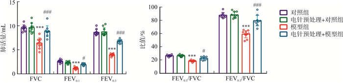
图 1 各组大鼠肺功能比较
注: 与对照组比较, ***P < 0.001;与模型组比较, #P < 0.05, ###P < 0.001。x±s, n=10。
Figure 1. Comparison of lung function of the rats in each group

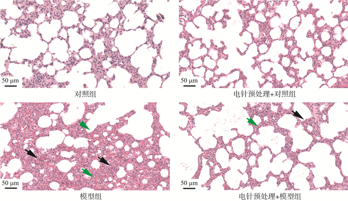
图 2 各组大鼠肺组织形态学比较(HE染色, ×200)
注: 黑色箭头指示为大量炎性细胞浸润; 绿色箭头指示为红细胞渗出
Figure 2. Comparison of lung histomorphology of rats in each group (HE staining, ×200)

图 3 各组大鼠肺组织W/D值比较
注: 与对照组比较, ***P < 0.001;与模型组比较, ##P < 0.01。x±s, n=10。
Figure 3. Comparison of wet/dry weight ratio of lung tissue of rats in each group

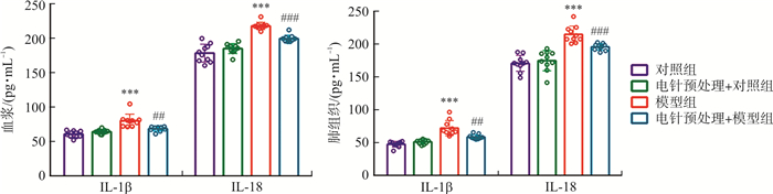
图 4 各组大鼠血浆及肺组织中IL-1β、IL-18含量比较
注: 与对照组比较, ***P < 0.001;与模型组比较, ##P < 0.01, ###P < 0.001。x±s, n=10。
Figure 4. Comparison of IL-1β and IL-18 in plasma and lung tissue of rats in each group

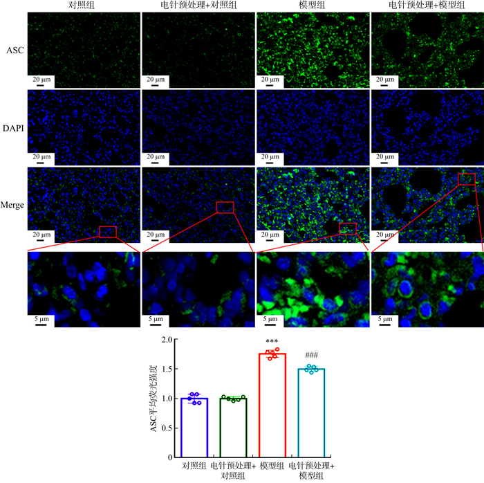
图 5 各组大鼠肺组织中ASC蛋白阳性表达比较(免疫荧光, ×400)
注: 细胞核呈蓝色, ASC蛋白阳性表达呈绿色。与对照组比较, ***P < 0.001;与模型组比较, ###P < 0.001。x±s, n=5。
Figure 5. Comparison of the positive expression of ASC protein in lung tissue of rats in each group (IF staining, ×400)
-
[1] LU QY, YU SF, MENG XY, et al. microRNAs: Important regulatory molecules in acute lung injury/acute respiratory distress syndrome[J]. Int J Mol Sci, 2022, 23(10): 5545. doi: 10.3390/ijms23105545 [2] LIU C, XIAO K, XIE LX. Advances in the use of exosomes for the treatment of ALI/ARDS[J]. Front Immunol, 2022, 13: 971189. doi: 10.3389/fimmu.2022.971189 [3] MEYER NJ, GATTINONI L, CALFEE CS. Acute respiratory distress syndrome[J]. Lancet, 2021, 398(10300): 622-637. doi: 10.1016/S0140-6736(21)00439-6 [4] 谢璨灿, 吴双华, 李峥嵘, 等. 电针刺激通过JAK1/STAT3通路减轻脓毒症大鼠的急性肺损伤[J]. 南方医科大学学报, 2020, 40(11): 1662-1667. doi: 10.12122/j.issn.1673-4254.2020.11.20XIE CC, WU SH, LI ZR, et al. Electroacupuncture protects septic rats from acute lung injury through the JAK1/STAT3 pathway[J]. J South Med Univ, 2020, 40(11): 1662-1667. doi: 10.12122/j.issn.1673-4254.2020.11.20 [5] LUO D, LIU L, ZHANG HM, et al. Electroacupuncture pretreatment exhibits lung protective and anti-inflammation effects in lipopolysaccharide-induced acute lung injury via SIRT1-dependent pathways[J]. Evid Based Complement Alternat Med, 2022, 2022: 2252218. [6] SHI XY, LI T, LIU YT, et al. HSF1 protects sepsis-induced acute lung injury by inhibiting NLRP3 inflammasome activation[J]. Front Immunol, 2022, 13: 781003. doi: 10.3389/fimmu.2022.781003 [7] WANG H, SUN XT, LU Q, et al. The mitochondrial redistribution of eNOS is involved in lipopolysaccharide induced inflammasome activation during acute lung injury[J]. Redox Biol, 2021, 41: 101878. doi: 10.1016/j.redox.2021.101878 [8] LI JM, BAI Y, TANG YT, et al. A 4-benzene-indol derivative alleviates LPS-induced acute lung injury through inhibiting the NLRP3 inflammasome[J]. Front Immunol, 2022, 13: 812164. doi: 10.3389/fimmu.2022.812164 [9] XU J, LU LQ, LI LF. NEK7: A novel promising therapy target for NLRP3-related inflammatory diseases[J]. Acta Biochim Biophys Sin, 2016, 48(10): 966-968. doi: 10.1093/abbs/gmw080 [10] SUN ZZ, GONG W, ZHANG Y, et al. Physiological and pathological roles of mammalian NEK7[J]. Front Physiol, 2020, 11: 606996. doi: 10.3389/fphys.2020.606996 [11] 刘瑞莲. 利多卡因通过P2X7R/NLRP3/Caspase-1途径对脂多糖诱导急性肺损伤大鼠的保护作用研究[D]. 广州: 广州医科大学, 2018.LIU RL. Protective effect of lidocaine on lipopolysaccharide induced acute lung injury in rat via attenuating P2X7R/NLRP3/caspase-1 pathway[D]. Guangzhou: Guangzhou Medical University, 2018. [12] 刘心月, 苏景超, 张新芳, 等. 电针预处理对脂多糖诱导的脓毒症急性肺损伤大鼠肺组织中血管紧张素转化酶2、血管紧张素(1-7) 的影响[J]. 针刺研究, 2022, 47(8): 684-689. https://www.cnki.com.cn/Article/CJFDTOTAL-XCYJ202208005.htmLIU XY, SU JC, ZHANG XF, et al. Electroacupuncture preconditioning improves pulmonary function via inhibiting inflammatory response and up-regulating expression of ACE2 and Ang (1-7) in lipopolysaccharide-induced acute lung injury rats[J]. Acupunct Res, 2022, 47(8): 684-689. https://www.cnki.com.cn/Article/CJFDTOTAL-XCYJ202208005.htm [13] 李忠仁. 实验针灸学[M]. 2版. 北京: 中国中医药出版社, 2007.LI ZR. Experimental Acupuncture[M]. 2nd ed. Beijing: China press of traditional Chinese medicine, 2007. [14] 杨英伟, 李建, 刘恩顺, 等. 204例ALI/ARDS患者中医证候分布与演变特征研究[J]. 中华中医药杂志, 2015, 30(3): 911-913.YANG YW, LI J, LIU ES, et al. Study on the TCM syndrome distribution and evolution characteristic of 204 ALI/ARDS patients[J]. China J Tradit Chin Med Pharm, 2015, 30(3): 911-913. [15] 徐弋茜, 崔翔, 刘坤, 等. 电针和预电针改善急性肺损伤大鼠肺功能的效应差异[J]. 针刺研究, 2022, 47(7): 580-586. doi: 10.13702/j.1000-0607.20211299XU YQ, CUI X, LIU K, et al. Comparison of effects of routine electroacupuncture and pre-electroacupuncture in improving lung function in acute lung injury rats[J]. Acupunct Res, 2022, 47(7): 580-586. doi: 10.13702/j.1000-0607.20211299 [16] LIU SB, WANG ZF, SU YS, et al. A neuroanatomical basis for electroacupuncture to drive the vagal-adrenal axis[J]. Nature, 2021, 598(7882): 641-645. doi: 10.1038/s41586-021-04001-4 [17] ZHANG YG, ZHENG L, DENG HM, et al. Electroacupuncture alleviates LPS-induced ARDS through α7 nicotinic acetylcholine receptor-mediated inhibition of ferroptosis[J]. Front Immunol, 2022, 13: 832432. doi: 10.3389/fimmu.2022.832432 [18] 张毅. 电针预处理调控ALI大鼠肺泡巨噬细胞M1极化的作用研究[D]. 合肥: 安徽中医药大学, 2021.ZHANG Y. Effect of electro-acupuncture pretreatment on M1 polarization of alveolar macrophages in acute lung injury rats[D]. Hefei: Anhui University of Chinese Medicine, 2021. [19] AVECILLAS JF, FREIRE AX, ARROLIGA AC. Clinical epidemiology of acute lung injury and acute respiratory distress syndrome: Incidence, diagnosis, and outcomes[J]. Clin Chest Med, 2006, 27(4): 549-557. doi: 10.1016/j.ccm.2006.06.001 [20] NOVICK D, KIM S, KAPLANSKI G, et al. Interleukin-18, more than a Th1 cytokine[J]. Semin Immunol, 2013, 25(6): 439-448. doi: 10.1016/j.smim.2013.10.014 [21] YANG J, YANG JW, HUANG XF, et al. Glibenclamide alleviates LPS-induced acute lung injury through NLRP3 inflammasome signaling pathway[J]. Mediators Inflamm, 2022, 2022: 8457010. [22] DUAN YH, WANG JH, CAI J, et al. The leucine-rich repeat (LRR) domain of NLRP3 is required for NLRP3 inflammasome activation in macrophages[J]. J Biol Chem, 2022, 298(12): 102717. doi: 10.1016/j.jbc.2022.102717 [23] SHAO BZ, XU ZQ, HAN BZ, et al. NLRP3 inflammasome and its inhibitors: A review[J]. Front Pharmacol, 2015, 6: 262. [24] SONG H, ZHAO CY, YU ZX, et al. UAF1 deubiquitinase complexes facilitate NLRP3 inflammasome activation by promoting NLRP3 expression[J]. Nat Commun, 2020, 11(1): 6042. doi: 10.1038/s41467-020-19939-8 [25] ZANGIABADI S, ABDUL-SATER AA. Regulation of the NLRP3 inflammasome by posttranslational modifications[J]. J Immunol, 2022, 208(2): 286-292. doi: 10.4049/jimmunol.2100734 [26] PAIK S, KIM JK, SILWAL P, et al. An update on the regulatory mechanisms of NLRP3 inflammasome activation[J]. Cell Mol Immunol, 2021, 18(5): 1141-1160. doi: 10.1038/s41423-021-00670-3 [27] GUAN XX, YANG HH, ZHONG WJ, et al. Fn14 exacerbates acute lung injury by activating the NLRP3 inflammasome in mice[J]. Mol Med, 2022, 28(1): 85. doi: 10.1186/s10020-022-00514-4 [28] ZHANG Y, LI XR, GRAILER JJ, et al. Melatonin alleviates acute lung injury through inhibiting the NLRP3 inflammasome[J]. J Pineal Res, 2016, 60(4): 405-414. doi: 10.1111/jpi.12322 [29] FRY AM, BAYLISS R, ROIG J. Mitotic regulation by NEK kinase networks[J]. Front Cell Dev Biol, 2017, 5: 102. doi: 10.3389/fcell.2017.00102 [30] SHARIF H, WANG L, WANG WL, et al. Structural mechanism for NEK7-licensed activation of NLRP3 inflammasome[J]. Nature, 2019, 570(7761): 338-343. doi: 10.1038/s41586-019-1295-z [31] HE Y, ZENG MY, YANG DH, et al. NEK7 is an essential mediator of NLRP3 activation downstream of potassium efflux[J]. Nature, 2016, 530(7590): 354-357. doi: 10.1038/nature16959 [32] WANG Y, ZENG Z, RAN JR, et al. The critical role of potassium efflux and Nek7 in Pasteurella multocida -induced NLRP3 inflammasome activation[J]. Front Microbiol, 2022, 13: 849482. doi: 10.3389/fmicb.2022.849482 [33] LIU H, GU CP, LIU MJ, et al. NEK7 mediated assembly and activation of NLRP3 inflammasome downstream of potassium efflux in ventilator-induced lung injury[J]. Biochem Pharmacol, 2020, 177: 113998. -

 下载:
下载:


 点击查看大图
计量
- 文章访问数: 159
- HTML全文浏览量: 22
- PDF下载量: 16
- 被引次数: 0
