

Eucommia Folium Extracts Alleviate Nonalcoholic Fatty Liver Disease in vivo and in vitro Based on the Network Pharmacology Study
doi: 10.14148/j.issn.1672-0482.2022.0703
-
摘要:
目的 基于网络药理学探讨杜仲叶提取物(Eucommia ulmoides leaf extracts, ELE)改善非酒精性脂肪肝(Nonalcoholic fatty liver disease, NAFLD)的有效成分、作用靶点及可能的作用机制。 方法 使用CNKI、万方、TCMSP、Pubmed等数据库、获取并筛选杜仲叶活性成分; 使用TCMSP数据库和SwissTargetPrediction数据库检索并预测ELE活性成分的靶点; 使用Genecard数据库和Drugbank数据库收集NAFLD疾病的靶点; 使用String 11.0构建蛋白互作(Protein-protein interaction)网络图, 分析蛋白靶点之间的关系; 使用Cytoscape 3.7.1软件构建化合物-疾病靶点-通路网络图, 分析三者之间相互作用的关系; 使用DAVID数据库, 对靶点进行GO分析和KEGG通路富集分析, 并对关键靶点进行实验验证。 结果 ELE改善NAFLD疾病靶点网络主要包含9个化合物和35个靶点, 与脂质代谢相关的靶点有PPARα和CPT-1A, 与NAFLD相关的通路主要有胰岛素抵抗、AMPK信号通路。体内、体外实验发现ELE能通过激活AMPKα及增强靶点PPARα和CPT-1A的表达, 增强脂质氧化, 改善NAFLD。 结论 ELE能增强脂质代谢, 改善NAFLD, 其机制主要与AMPK信号通路相关。 Abstract:OBJECTIVE To explore the active ingredients, targets and possible mechanism of ELE alleviating NAFLD based on the method of network pharmacology. METHODS The components in ELE were found in CNKI, Wanfang, TCMSP and Pubmed database. The components targets were found in TCMSP and swisstarget prediction database. NAFLD targets were found in genecard database and drugbank database. PPI network was established to analyze the interaction among targets by String 11.0 database. Compounds-targets-pathways network was established by the software of Cytoscape. Go and KEGG analysis were carried by david database. In addition the key targets were verified by cell and animals experiments. RESULTS The network of ELE against NAFLD contains 9 compounds and 35 targets. The targets related to lipid metabolism are PPARα and CPT-1A, and the signaling pathways mainly related to lipid metabolism are insulin resistance and AMPK signaling pathway. In vivo and in vitro experiments indicated that ELE could enhance lipid oxidation and improve NAFLD by activating AMPKα and enhancing the expression of PPARα and CPT-1A. CONCLUSION ELE can enhance lipid metabolism and improve NAFLD mainly related to AMPK signaling pathway. -
Key words:
- network pharmacology /
- Eucommia ulmoides leaf extract /
- NAFLD /
- AMPK signaling pathway
-

Figure 1. Protein-protein interaction (PPI) network
Note: (A) Intersection gene of ELE targets and NAFLD related targets. ELE consists 346 targets generated from 9 active compounds. NAFLD consists 180 targets and there are 35 common targets of ELE and NAFLD. (B) Core network (PPI) diagram of ELE and NAFLD. The nodes get larger with increasing degree. Edges: PPIs between putative targets of ELE and their interactive partners.

Figure 2. The compound-compound targets-pathway network of ELE
Note: active compounds in blue, target in pink, pathway in red

Figure 3. Gene ontology and pathway enrichment analysis of identified related targets
Note: (A) biological process. (B) molecular function. (C) cellular component. (D) pathway analysis.

Figure 4. The effect of palmitic acid on cell viability of L02 and HepG2 cells
Note: L02 and HepG2 cells were treated with palmitic acid (200 μmol·L-1, 250 μmol·L-1 and 300 μmol·L-1, respectively.) for 24 h. (A) L02 cell viability was detected by MTT assay (n=6). (B) HepG2 cell viability was detected by MTT assay (n=6).

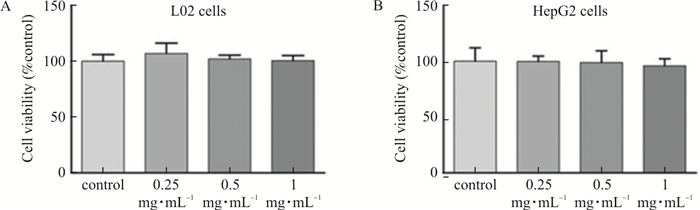
Figure 5. The effect of ELE on cell viability of L02 and HepG2 cells
Note: L02 and HepG2 cells were treated with ELE (0.25 mg·mL-1, 0.5 mg·mL-1 and 1 mg·mL-1, respectively.) for 24 h. (A) L02 cell viability was detected by MTT assay. (B) HepG2 cell viability was detected by MTT assay. x±s, n=6.

Figure 6. ELE alleviates lipid accumulation in PA-treated L02 and HepG2 cells
Note: L02 and HepG2 cells were pretreated with 200 μmol·L-1 PA for 24 h and treated with 0.25 mg·mL-1, 0.5 mg·mL-1, and 1 mg·mL-1 ELE for an additional 24 h. (A) Oil red O staining images of L02 cells (magnification 200×). Photographs are representative images of three independent experiments. (B) Quantitative oil red O contents measured at 510 nm. (C~D) Intracellular TG, TC levels measured using commercial kit. (n=6). (E) Oil red O staining images of HepG2 cells (magnification 200×). Photographs are representative images of three independent experiments. (F) Quantitative Oil red O contents measured at 510 nm. G-H. Intracellular TG, TC levels measured using acommercial kit. x±s, n=6.

Figure 7. ELE alleviates lipid metabolism by activating AMPKα in PA-treated L02 and HepG2 Cells
Note: L02 and HepG2 cells were pretreated with 200 μmol·L-1 PA for 24 h and treated with 0.25 mg·mL-1, 0.5 mg·mL-1, and 1 mg·mL-1 ELE for an additional 24 h. (A) The protein expression of P-AMPKα, AMPKα, PPARα, CPT-1A in PA-treated L02 cells (n=3). (B) Densitometric analysis of western blot. (C-D) The gene expression of CPT-1A, PPARα in PA-treated L02 cells (n=3). (E) The protein expression of P-AMPKα, AMPKα, PPARα, CPT-1A in PA-treated HepG2 cells (n=3). (F) Densitometric analysis of western blot in (E). (G-H) Gene expression of CPT-1A, PPARα in PA-treated HepG2 cells (n=3).

Figure 8. ELE attenuates liver obesity and injury in HFD-fed mice
Note: With the HFD, mice were treated daily with ELE (low dosage group: 1 g·kg-1, high dosage group: 2 g·kg-1) or PBS (control group, model group). (A) Macroscopic view of mice liver in different groups (n=3). (B) Effect of ELE on mice liver weight (n=7-8). (C) Effect of ELE on the ratios of liver weight to body weight (n=7-8). (D) The changes of mice body weight (n=7-8). (E) Effect of ELE on belly weight (n=7-8). (F) Effect of ELE on serum ALT (n=7-8). (G) Effect of ELE on serum AST (n=7-8).

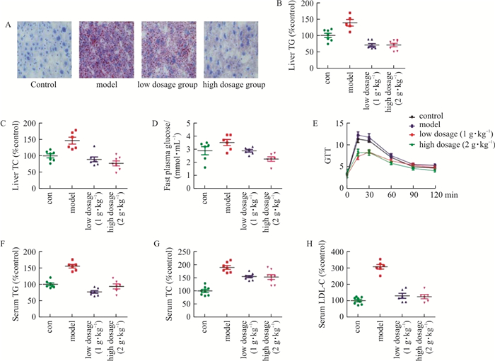
Figure 9. ELE ameliorates hepatic steatosis in HFD-fed mice
Note: With the HFD, mice were treated daily with ELE (low dosage group: 1 g·kg-1, high dosage group: 2 g·kg-1) or PBS (control group, model group). (A) Oil red O staining of liver tissue (magnification 400×) (n=3). (B-C) Liver TG, TC levels measured using a commercial kit (n=7-8). (D) The level of fast blood glucose (n=7-8). (E) The level of oral glucose tolerance (n=7-8). (F-H) Serum TG, TC, LDL-C levels measured using a commercial kit (n=7-8).

Figure 10. ELE alleviates liver lipid metabolism by activating AMPKα in HFD-fed mice
Note: With the HFD, mice were treated daily with ELE (low dosage group: 1 g·kg-1, high dosage group: 2 g·kg-1) or PBS (control group, model group). (A) Protein expression of P-AMPKα, AMPKα, PPARα, CPT-1A in HFD-fed mice liver tissue. (B) Densitometric analysis of western blot (n=3). (C-D) The gene expression of CPT-1A, PPARα in HFD-fed mice liver tissue (n=3).
Table 1. Screening of active componets of Eucommia folium
Mol-ID Ingredient Chemical structure Molecular formula OB(≥30%) DL(≥0.18) MOL000422 Kaempferol 
C15H10O6 41.88 0.24 MOL000098 Quercetin 
C15H10O7 46.43 0.28 MOL000006 Luteolin 
C15H10O6 36.16 0.25 MOL002714 Baicalein 
C15H10O5 33.52 0.21 MOL001668 Geniposidic acid 
C16H22O10 19.59 0.41 MOL002813 Aucubin 
C15H22O9 35.56 0.33 MOL001955 Chlorogenic acid 
C16H18O9 11.93 0.33 MOL000497 Licochalcone A 
C21H22O4 40.79 0.29 MOL002928 Oroxylin A 
C16H12O5 41.37 0.23  下载: 导出CSV
Table 2. The main targets of Eucommia folium in the treatment of NAFLD
Gene symbol Gene name Degree MPO Myeloperoxidase 4 MMP2 Matrix metalloproteinase 2 9 MMP9 Matrix metalloproteinase 9 11 MMP1 Matrix metalloproteinase 1 4 IKBKB Inhibitor of nuclear factor kappa B kinase beta subunit 10 ELANE Leukocyte elastase 1 ADORA2A Adenosine A2a receptor 7 NOS2 Nitric oxide synthase, inducible (by homology) 5 ACE Angiotensin-converting enzyme 3 TTR Transthyretin 5 INSR Insulin receptor 10 IGF1R Insulin-like growth factor I receptor 6 APP Beta amyloid A4 protein 9 CFTR Cystic fibrosis transmembrane conductance regulator 5 FASN Fatty acid synthase 2 CPT-1A Carnitine Palmitoyltransferase 1A 3 PPARA Peroxisome proliferator-activated receptor alpha 3 ADORA1 Adenosine A1 receptor (by homology) 7 NR1H4 Bile acid receptor FXR 1 AKT1 Serine/threonine-protein kinase AKT 13 EGFR Epidermal growth factor receptor erbB1 12 PIK3CA PI3-kinase p110-alpha subunit 10 PDGFRB Platelet-derived growth factor receptor beta 3 VCP Transitional endoplasmic reticulum ATPase 1 PIK3R1 PIK3R1 14 ESR1 Estrogen receptor alpha 5 TERT Telomerase reverse transcriptase 4 PTPN11 Protein-tyrosine phosphatase 2C 4 STAT3 Signal transducer and activator of transcription 3 8 MAPK8 c-Jun N-terminal kinase 1 8 CASP8 Caspase-8 6 SIRT1 NAD-dependent deacetylase sirtuin 1 3 F2 Thrombin 4
下载: 导出CSV
Table 3. Information of signaling pathways (10 of 77)
ID Pathway Degree Count P Value hsa05200 Pathways in cancer 15 16 6.981 06E-11 hsa05205 Proteoglycans in cancer 10 11 1.781 88E-08 hsa04014 Ras signaling pathway 10 11 5.728 88E-08 hsa04931 Insulin resistance 9 10 1.166 26E-09 hsa04068 Foxo signaling pathway 9 10 8.006 91E-09 hsa04152 AMPK signaling pathway 8 9 7.939 08E-08 hsa05161 Hepatitis B 9 9 2.856 03E-07 hsa04932 Non-alcoholic fatty liver disease (NAFLD) 10 9 3.904 25E-07 hsa05212 Pancreatic cancer 8 8 1.701 12E-08 hsa04066 HIF-1 signaling pathway 7 8 2.626 42E-07
下载: 导出CSV
Table 4. Information of biological process (10 of 132)
ID Term Count P Value GO: 0043066 negative regulation of apoptotic process 15 8.01E-14 GO: 0048015 phosphatidylinositol-mediated signaling 7 6.39E-08 GO: 0014066 regulation of phosphatidylinositol 3-kinase signaling 6 4.76E-07 GO: 0044267 cellular protein metabolic process 6 3.72E-06 GO: 0050900 leukocyte migration 6 4.38E-06 GO: 0001934 positive regulation of protein phosphorylation 6 5.34E-06 GO: 0010629 negative regulation of gene expression 6 7.73E-06 GO: 0040014 regulation of multicellular organism growth 4 1.91E-05 GO: 0046326 positive regulation of glucose import 4 2.97E-05 GO: 0032355 response to estradiol 5 3.30E-05
下载: 导出CSV
Table 5. Information of molecular function (10 of 42)
ID Term Count P Value GO: 0005515 protein binding 31 8.612 92E-06 GO: 0042802 identical protein binding 14 9.111 58E-10 GO: 0019899 enzyme binding 9 2.447 57E-07 GO: 0043560 insulin receptor substrate binding 5 4.479 75E-09 GO: 0046934 phosphatidylinositol-4, 5-bisphosphate 3-kinase activity 5 7.042 79E-06 GO: 0043548 phosphatidylinositol 3-kinase binding 4 7.075 77E-06 GO: 0004879 RNA polymerase Ⅱ transcription factor activity, ligand-activated sequence-specific DNA binding 4 5.093 10E-05 GO: 0004175 endopeptidase activity 4 0.000 172 604 GO: 0043559 insulin binding 3 3.92261E-05 GO: 0030235 nitric-oxide synthase regulator activity 3 0.000109417
下载: 导出CSV
Table 6. Information of cellular component (10 of 23)
ID Term Count P Value GO: 0005886 plasma membrane 19 0.000 112 612 GO: 0005737 cytoplasm 19 0.002 464 041 GO: 0005634 nucleus 18 0.010 172 407 GO: 0005829 cytosol 15 0.001 507 867 GO: 0016020 membrane 12 0.001 559 922 GO: 0005739 mitochondrion 10 0.000 567 226 GO: 0005615 extracellular space 9 0.002 795 184 GO: 0045121 membrane raft 5 0.000 563 769 GO: 0043235 receptor complex 4 0.001 689 032 GO: 0005943 phosphatidylinositol 3-kinase complex, class IA 2 0.003 727 965
下载: 导出CSV
-
[1] LONARDO A, TARGHER G. NAFLD in the 20's. from epidemiology to pathogenesis and management of nonalcoholic fatty liver disease[J]. Curr Pharm Des, 2020, 26(10): 991-992. doi: 10.2174/138161282610200424091752 [2] COLLIER J. Non-alcoholic fatty liver disease[J]. Medicine, 2007, 35(2): 86-88. doi: 10.1016/j.mpmed.2006.11.010 [3] COBBINA E, AKHLAGHI F. Non-alcoholic fatty liver disease (NAFLD) — pathogenesis, classification, and effect on drug metabolizing enzymes and transporters[J]. Drug Metab Rev, 2017, 49(2): 197-211. doi: 10.1080/03602532.2017.1293683 [4] PAVLIDES M, COBBOLD JFL. Non-alcoholic fatty liver disease[J]. Medicine, 2015, 43(10): 585-589. doi: 10.1016/j.mpmed.2015.07.004 [5] KEI N. Multidisciplinary pharmacotherapeutic options for nonalcoholic fatty liver disease[J]. Int J Hepatol, 2012, 2012: 950693. [6] CALDWELL S. NASH Therapy: Omega 3 supplementation, vitamin E, insulin sensitizers and statin drugs[J]. Clin Mol Hepatol, 2017, 23(2): 103-108. doi: 10.3350/cmh.2017.0103 [7] VERGANI L. Fatty acids and effects on in vitro and in vivo models of liver steatosis[J]. Curr Med Chem, 2019, 26(19): 3439-3456. doi: 10.2174/0929867324666170518101334 [8] STEFANOVIC-RACIC M, PERDOMO G, MANTELL BS, et al. A moderate increase in carnitine palmitoyltransferase 1a activity is sufficient to substantially reduce hepatic triglyceride levels[J]. Am J Physiol Endocrinol Metab, 2008, 294(5): E969-E977. doi: 10.1152/ajpendo.00497.2007 [9] DUVAL C, MULLER M, KERSTEN S. PPARα and dyslipidemia[J]. Biochim Biophys Acta BBA Mol Cell Biol Lipids, 2007, 1771(8): 961-971. [10] MONTAGNER A, POLIZZI A, FOUCHE E, et al. Liver PPARα is crucial for whole-body fatty acid homeostasis and is protective against NAFLD[J]. Gut, 2016, 65(7): 1202-1214. doi: 10.1136/gutjnl-2015-310798 [11] JUNG YH, AGRAWAL GK, RAKWAL R, et al. Functional characterization of OsRacB GTPase — a potentially negative regulator of basal disease resistance in rice[J]. Plant Physiol Biochem, 2006, 44(1): 68-77. doi: 10.1016/j.plaphy.2005.12.001 [12] LEE GH, LEE HY, PARK SA, et al. Eucommia ulmoides leaf extract ameliorates steatosis induced by high-fat diet in rats by increasing lysosomal function[J]. Nutrients, 2019, 11(2): 426. doi: 10.3390/nu11020426 [13] LEE HY, LEE GH, YOON Y, et al. Rhus verniciflua and Eucommia ulmoides protects against high-fat diet-induced hepatic steatosis by enhancing anti-oxidation and AMPK activation[J]. Am J Chin Med, 2019, 47(6): 1253-1270. doi: 10.1142/S0192415X19500642 [14] HAO DC, XIAO PG. Network pharmacology: A Rosetta Stone for traditional Chinese medicine[J]. Drug Dev Res, 2014, 75(5): 299-312. doi: 10.1002/ddr.21214 [15] RU JL, LI P, WANG JN, et al. TCMSP: A database of systems pharmacology for drug discovery from herbal medicines[J]. J Cheminform, 2014, 6: 13. doi: 10.1186/1758-2946-6-13 [16] KIM S. Getting the most out of PubChem for virtual screening[J]. Expert Opin Drug Discov, 2016, 11(9): 843-855. doi: 10.1080/17460441.2016.1216967 [17] SAFRAN M, CHALIFA-CASPI V, SHMUELI O, et al. Human gene-centric databases at the Weizmann institute of science: GeneCards, UDB, CroW 21 and HORDE[J]. Nucleic Acids Res, 2003, 31(1): 142-146. doi: 10.1093/nar/gkg050 [18] SZKLARCZYK D, FRANCESCHINI A, WYDER S, et al. STRING v10: Protein-protein interaction networks, integrated over the tree of life[J]. Nucleic Acids Res, 2014, 43(D1): D447-D452. [19] HUANG DW, SHERMAN BT, TAN QN, et al. DAVID Bioinformatics Resources: Expanded annotation database and novel algorithms to better extract biology from large gene lists[J]. Nucleic Acids Res, 2007, 35(suppl_2): W169-W175. doi: 10.1093/nar/gkm415 [20] LIU C, GUO FF, XIAO JP, et al. Research progress on chemical constituents and pharmacological effects of different parts of Eucommia ulmoides[J]. China J Chin Mater Med, 2020, 45(3): 497-512. [21] HAO S, XIAO Y, LIN Y, et al. Chlorogenic acid-enriched extract from Eucommia ulmoides leaves inhibits hepatic lipid accumulation through regulation of cholesterol metabolism in HepG2 cells[J]. Pharm Biol, 2016, 54(2): 251-259. doi: 10.3109/13880209.2015.1029054 [22] ABDELMALEK MF, DIEHL AM. Nonalcoholic fatty liver disease as a complication of insulin resistance[J]. Med Clin N Am, 2007, 91(6): 1125-1149. doi: 10.1016/j.mcna.2007.06.001 [23] HERNANDEZ-RODAS MC, VALENZUELA R, VIDELA LA. Relevant aspects of nutritional and dietary interventions in non-alcoholic fatty liver disease[J]. Int J Mol Sci, 2015, 16(10): 25168-25198. doi: 10.3390/ijms161025168 [24] YU JH, XIAO ZC, ZHAO RZ, et al. Paeoniflorin suppressed IL-22 via p38 MAPK pathway and exerts anti-psoriatic effect[J]. Life Sci, 2017, 180: 17-22. doi: 10.1016/j.lfs.2017.04.019 [25] HOPKINS AL. Network pharmacology: The next paradigm in drug discovery[J]. Nat Chem Biol, 2008, 4(11): 682-690. doi: 10.1038/nchembio.118 [26] LAU JKC, ZHANG X, YU J. Animal models of non-alcoholic fatty liver disease: Current perspectives and recent advances[J]. J Pathol, 2017, 241(1): 36-44. doi: 10.1002/path.4829 [27] WANG CH, TAO QM, WANG XH, et al. Impact of high-fat diet on liver genes expression profiles in mice model of nonalcoholic fatty liver disease[J]. Environ Toxicol Pharmacol, 2016, 45: 52-62. doi: 10.1016/j.etap.2016.05.014 [28] SUN X, DUAN XP, WANG CY, et al. Protective effects of glycyrrhizic acid against non-alcoholic fatty liver disease in mice[J]. Eur J Pharmacol, 2017, 806: 75-82. [29] MI Y, TAN DH, HE Y, et al. Melatonin modulates lipid metabolism in HepG2 cells cultured in high concentrations of oleic acid: AMPK pathway activation may play an important role[J]. Cell Biochem Biophys, 2018, 76(4): 463-470. [30] FANG K, WU F, CHEN G, et al. Diosgenin ameliorates palmitic acid-induced lipid accumulation via AMPK/ACC/CPT-1A and SREBP-1c/FAS signaling pathways in LO2 cells[J]. BMC Complement Altern Med, 2019, 19(1): 255. [31] MOODY L, XU GB, CHEN H, et al. Epigenetic regulation of carnitine palmitoyltransferase 1 (Cpt1a) by high fat diet[J]. Biochim Biophys Acta BBA Gene Regul Mech, 2019, 1862(2): 141-152. [32] PAWLAK M, LEFEBVRE P, STAELS B. Molecular mechanism of PPARα action and its impact on lipid metabolism, inflammation and fibrosis in non-alcoholic fatty liver disease[J]. J Hepatol, 2015, 62(3): 720-733. -


 点击查看大图

计量
- 文章访问数: 296
- HTML全文浏览量: 100
- PDF下载量: 45
- 被引次数: 0
