

Molecular Mechanism Research of Wenshen Yanggan Decoction on Dopaminergic Neuron Injury in MPTP-Induced Parkinson's Disease
-
摘要:
目的 探讨温肾养肝汤保护多巴胺(Dopamine, DA)能神经元, 延缓帕金森病(Parkinson's disease, PD)进程的作用机制。 方法 将50只小鼠随机分为正常组, 模型组, 温肾养肝汤高、低剂量组, 金刚烷胺组, 每组10只, 除正常组外, 其余组小鼠腹腔注射30 mg·kg-1 1-甲基-4-苯基-1, 2, 3, 6-四氢吡啶(MPTP), 每周2次, 连续4周, 建立MPTP致小鼠PD模型, 灌胃温肾养肝汤,每日1次,连续3周。观察PD小鼠的行为学变化;免疫组化和尼氏染色法检测DA能神经元细胞数;HPLC法检测病理组织中DA、3, 4-二羟基苯乙酸(3, 4-Dihydroxyphenylacetic acid, DOPAC)和高香草酸(Homovanillic acid, HVA)含量;比色法测定小鼠黑质线粒体中ATP酶(ATPase)、线粒体呼吸链复合物Ⅰ(ComplexⅠ)活性;Western blot测定PINK1、Parkin、VDAC1、LC3Ⅱ/Ⅰ、P62蛋白相对表达量。 结果 行为学检测显示, 与模型组相比, 温肾养肝汤能明显改善小鼠的肌肉力量和运动平衡(P < 0.01), 延长小鼠在滚筒上的时间(P < 0.01);免疫组化显示, 温肾养肝汤高剂量组可观察到大量酪氨酸氢化酶(Tyrosine hydrogenase,TH)免疫反应呈阳性的细胞, 明显的细胞突起, 与尼氏染色结果一致, 同时小鼠纹状体中DA和HVA含量明显提高(P < 0.05, P < 0.01);黑质线粒体酶活性和自噬通路相关蛋白研究显示, 高剂量温肾养肝汤能明显提高ATPase和ComplexⅠ活性(P < 0.01), 同时还能上调模型小鼠的PINK1(P < 0.05)、Parkin(P < 0.01)、LC3Ⅱ/Ⅰ(P < 0.01)蛋白表达, 抑制P62蛋白表达(P < 0.01)。 结论 温肾养肝汤通过提高线粒体功能, 激活PINK1/Parkin介导的线粒体自噬过程来保护DA能神经元, 改善PD模型小鼠行为学功能障碍, 延缓PD进程。 -
关键词:
- 温肾养肝汤 /
- 帕金森病 /
- 多巴胺能神经元 /
- 线粒体自噬 /
- PINK1/Parkin
Abstract:OBJECTIVE To explore the potential mechanism of Wenshen Yanggan Decoction on protecting dopaminergic neurons and delaying the process of Parkinson's disease (PD). METHODS Fifty mice were randomly divided into 5 groups (n=10): control group, model group, Wenshen Yanggan Decoction high dose group, Wenshen Yanggan Decoction low dose group and amantadine group. Except for the control group, mice were injected intraperitoneally with 30 mg · kg-1 of 1-methyl-4-phenyl-1, 2, 3, 6-tetrahydropyridine (MPTP) twice a week for 4 consecutive weeks to establish a Parkinson's disease (PD) mouse model. The mice were administrated with Wenshen Yanggan Decoction by gavage once a day for 3 weeks. The behavioral effects of mice were observed. Immunohistochemistry and Nissl staining were used to detect the number of DA neurons. The contents of DA, 3, 4-dihydroxyphenylacetic acid (DOPAC) and homovanillic acid (HVA) in pathological tissues were measured by HPLC. Colorimetric method was used to determine the activities of ATPase and mitochondrial respiratory chain ComplexⅠ in the substantia nigra. The protein expression levels of PINK1, Parkin, VDAC1, LC3Ⅱ/Ⅰ, and P62 were detected by Western blot. RESULTS The behavioral tests showed that compared with the model group, Wenshen Yanggan Decoction significantly improved the muscle strength and exercise balance of mice (P < 0.01), and prolonged the duration of mice on the roller (P < 0.01). The Results of the immunohistochemistry demonstrated that a large number of TH immunoreactive cells and obvious cell protrusions were observed in Wenshen Yanggan Decoction high dose group, which was consistent with the Results of the Nissl staining assay. In addition, Wenshen Yanggan Decoction significantly increased the levels of DA and HVA (P < 0.05, P < 0.01) in the striatum. Furthermore, treatment with the high dose Wenshen Yanggan Decoction profoundly promoted the activities of ATPase and ComplexⅠ(P < 0.01), up-regulated the protein levels of PINK1 (P < 0.05), Parkin (P < 0.01) and LC3Ⅱ/Ⅰ (P < 0.01), and inhibited P62 protein expression (P < 0.01) in MPTP-induced mice. CONCLUSION Wenshen Yanggan Decoction can improve mitochondrial function, and provide neuroprotective effect in PD by activating PINK1/Parkin mediated mitochondrial autophagy pathway. -

图 1 温肾养肝汤对PD小鼠运动障碍的影响
注:与正常组比较, **P < 0.01;与模型组比较, #P < 0.05, ##P < 0.01。x±s, n=10。
Figure 1. Effects of Wenshen Yanggan Decoction on dyskinesia in PD mice

图 2 温肾养肝汤对PD小鼠DA能神经元的影响
注:与正常组比较, **P < 0.01;与模型组比较, ##P < 0.01。x±s, n=3。
Figure 2. Effects of Wenshen Yanggan Decoction on dopaminergic neurons in PD mice

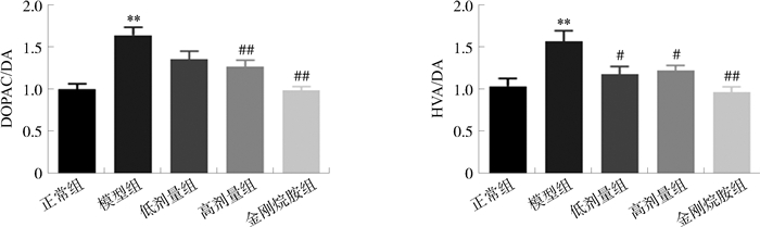
图 3 温肾养肝汤对PD小鼠纹状体DOPAC/DA和HVA/DA的影响
注:与正常组比较, **P < 0.01;与模型组比较, #P < 0.05, ##P < 0.01。x±s, n=6。
Figure 3. Effects of Wenshen Yanggan Decoction on DOPAC/DA and HVA/DA in PD mice

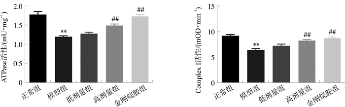
图 4 温肾养肝汤对PD小鼠线粒体酶活性的影响
注:与正常组比较, **P < 0.01;与模型组比较, ##P < 0.01。x±s, n=6。
Figure 4. Effects of Wenshen Yanggan Decoction on ATPase and ComplexⅠ activities in PD mice

图 5 温肾养肝汤对PD小鼠线粒体自噬通路蛋白表达的影响
注:与正常组比较, **P < 0.01;与模型组比较, #P < 0.05, ##P < 0.01。
Figure 5. Effects of Wenshen Yanggan Decoction on mitochondrial autophagy pathway-related protein expressions in PD mice
表 1 温肾养肝汤对PD小鼠纹状体DA及其代谢物的影响(x±s, ng·mg-1,n=6)
Table 1. Effects of Wenshen Yanggan Decoction on DA and its metabolites in PD mice (x±s, ng·mg-1, n=6)
组4别 DA DOPAC HVA 正常组 6.10±0.59 6.02±0.63 6.18±0.71 模型组 2.83±0.56** 4.51±0.37** 4.26±0.23** 低剂量组 3.95±0.51# 5.26±0.41 4.54±0.47 高剂量组 4.24±0.37## 5.30±0.49 5.11±0.19# 金刚烷胺组 5.55±0.63## 5.46±0.74# 5.31±0.62## 注:与正常组比较, **P < 0.01;与模型组比较, #P < 0.05, ##P < 0.01。  下载: 导出CSV
-
[1] OBESO JA, STAMELOU M, GOETZ CG, et al. Past, present, and future of Parkinson's disease: A special essay on the 200th Anniversary of the Shaking Palsy[J]. Mov Disord, 2017, 32(9): 1264-1310. doi: 10.1002/mds.27115 [2] DORSEY ER, SHERER T, OKUN MS, et al. The emerging evidence of the parkinson pandemic[J]. J Parkinsons Dis, 2018, 8(S1): S3-S8. doi: 10.3233/JPD-181474 [3] LI G, MA JF, CUI SS, et al. Parkinson's disease in China: A forty-year growing track of bedside work[J]. Transl Neurodegener, 2019, 8: 22. doi: 10.1186/s40035-019-0162-z [4] MENZIES FM, FLEMING A, RUBINSZTEIN DC. Compromised autophagy and neurodegenerative diseases[J]. Nat Rev Neurosci, 2015, 16(6): 345-357. doi: 10.1038/nrn3961 [5] BOSE A, BEAL MF. Mitochondrial dysfunction in Parkinson's disease[J]. J Neurochem, 2016, 139(14): 216-231. [6] 甘雪, 刘书一, 王正波. 线粒体自噬及功能障碍与帕金森病[J]. 中国比较医学杂志, 2020, 30(10): 121-127. doi: 10.3969/j.issn.1671-7856.2020.10.018GAN X, LIU SY, WANG ZB. Mitochondrial autophagy and dysfunction in Parkinson's disease[J]. Chin J Comp Med, 2020, 30(10): 121-127. doi: 10.3969/j.issn.1671-7856.2020.10.018 [7] 王苏雷, 杨卉, 陆艳, 等. 赵杨教授治疗帕金森病失眠经验及验案举隅[J]. 四川中医, 2018, 36(7): 17-19.WANG SL, YANG H, LU Y, et al. Professor Zhao Yang's experience in treating insomnia of Parkinson's disease[J]. J Sichuan Tradit Chin Med, 2018, 36(7): 17-19. [8] 陆艳, 张亚杰, 阮杰, 等. 肉苁蓉颗粒剂对帕金森病大鼠模型黑质纹状体多巴胺能神经元的保护作用研究[J]. 中华中医药学刊, 2016, 34(12): 2927-2931.LU Y, ZHANG YJ, RUAN J, et al. Nigrostriatal dopaminergic protection of Cistanche granule on rat model of Parkinson's disease[J]. Chin Arch Tradit Chin Med, 2016, 34(12): 2927-2931. [9] 赵伟, 孙国志. 不同种实验动物间用药量换算[J]. 畜牧兽医科技信息, 2010(5): 52-53.ZHAO W, SUN GZ. Conversion of drug dosage between different experimental animals[J]. Chin J Animal Husb Vet Med, 2010(5): 52-53. [10] HU M, LI FM, WANG WD. Vitexin protects dopaminergic neurons in MPTP-induced Parkinson's disease through PI3K/Akt signaling pathway[J]. Drug Des Devel Ther, 2018, 12: 565-573. doi: 10.2147/DDDT.S156920 [11] HASEGAWA K, YASUDA T, SHIRAISHI C, et al. Promotion of mitochondrial biogenesis by necdin protects neurons against mitochondrial insults[J]. Nat Commun, 2016, 7: 10943. doi: 10.1038/ncomms10943 [12] GENG XC, TIAN XF, TU PF, et al. Neuroprotective effects of echinacoside in the mouse MPTP model of Parkinson's disease[J]. Eur J Pharmacol, 2007, 564(1/2/3): 66-74. [13] 马浩洁. 基于PINK1基因探讨大补阴丸合牵正散对帕金森细胞模型线粒体的保护机制[D]. 北京: 北京中医药大学, 2018.MA HJ. The protective mechanism investigation of Dabuyin Pill combined with Qianzheng San on mitochondria in Parkinson's disease model cells based on PINK1 gene[D]. Beijing: Beijing University of Chinese Medicine, 2018. [14] 蒙健林, 梁健芬, 张兴博, 等. 帕金森病实验动物模型的研究进展及评价[J]. 中国实验动物学报, 2021, 29(3): 399-404. doi: 10.3969/j.issn.1005-4847.2021.03.016MENG JL, LIANG JF, ZHANG XB, et al. Research progress and evaluation on animal models of Parkinson's disease[J]. Acta Lab Animalis Sci Sin, 2021, 29(3): 399-404. doi: 10.3969/j.issn.1005-4847.2021.03.016 [15] CARTIER EA, PARRA LA, BAUST TB, et al. A biochemical and functional protein complex involving dopamine synthesis and transport into synaptic vesicles[J]. J Biol Chem, 2010, 285(3): 1957-1966. doi: 10.1074/jbc.M109.054510 [16] 李义. 帕金森氏病发病过程中多巴胺反应性氧化产物的蛋白修饰作用研究[D]. 遵义: 遵义医科大学, 2020.LI Y. The protein modification by reactive oxidation product of dopamine in the pathogenesis of Parkinson's disease[D]. Zunyi: Zunyi Medical College, 2020. [17] YOULE RJ, VAN DER BLIEK AM. Mitochondrial fission, fusion, and stress[J]. Science, 2012, 337(6098): 1062-1065. doi: 10.1126/science.1219855 [18] MALPARTIDA AB, WILLIAMSON M, NARENDRA DP, et al. Mitochondrial dysfunction and mitophagy in Parkinson's disease: From mechanism to therapy[J]. Trends Biochem Sci, 2021, 46(4): 329-343. doi: 10.1016/j.tibs.2020.11.007 [19] GEISLER S, HOLMSTRÖM KM, SKUJAT D, et al. PINK1/Parkin-mediated mitophagy is dependent on VDAC1 and p62/SQSTM1[J]. Nat Cell Biol, 2010, 12(2): 119-131. doi: 10.1038/ncb2012 [20] MA SF, ATTARWALA IY, XIE XQ. SQSTM1/p62: A potential target for neurodegenerative disease[J]. ACS Chem Neurosci, 2019, 10(5): 2094-2114. doi: 10.1021/acschemneuro.8b00516 -


 点击查看大图

计量
- 文章访问数: 176
- HTML全文浏览量: 73
- PDF下载量: 14
- 被引次数: 0
