

Effects of Electroacupuncture on Expression of Autophagy Related Proteins LC3-Ⅱ and Beclin1 in Rats after Cerebral Ischemia-Reperfusion
-
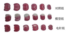
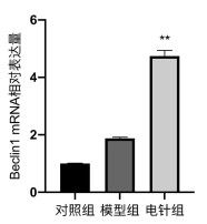
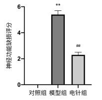
摘要: 目的 观察电针对脑缺血再灌注损伤大鼠神经功能的改善程度及自噬相关蛋白LC3-Ⅱ、Beclin1表达的影响。方法 根据随机数字表法,将72只雄性SD大鼠随机分为对照组、模型组和电针组,每组24只。采用Longa线栓法建立改良的急性局灶性脑缺血再灌注大鼠模型,并在缺血1 h后进行再灌注。参照改良的Zea Longa 8级神经功能缺损评分法对3组大鼠脑神经功能损伤程度进行评估。电针组于造模成功后5 min和16 h进行电针干预。于再灌注后24 h取材。TTC染色法观察各组大鼠脑梗死体积;HE染色法观察大鼠脑组织病理学变化;Western blot检测大鼠右侧纹状体脑组织中LC3-Ⅱ、Beclin1蛋白表达,qPCR检测Beclin1 mRNA表达。结果 对照组无行为学改变,模型组神经功能缺损评分显著高于对照组(P < 0.05);与模型组比较,电针组神经功能缺损评分明显低于模型组(P < 0.05)。电针组脑梗死体积较模型组明显减少(P < 0.05)。与模型组比较,电针组LC3-Ⅱ、Beclin1蛋白表达明显增加(P < 0.05),Beclin1 mRNA的表达亦明显增加(P < 0.05)。结论 电针治疗可能通过促进自噬相关蛋白LC3-Ⅱ、Beclin1的表达,调控自噬的发生,减小大鼠脑梗死体积,改善神经功能缺损,实现对脑缺血再灌注损伤大鼠的保护效应。Abstract: OBJECTIVE To observe the improvement degree of nerve function and the effect of electroacupuncture on autophagy related proteins LC3-Ⅱ and Beclin1 expression in rats with cerebral ischemia-reperfusion injury.METHODS According to the random number table method, 72 male SD rats were randomly divided into control group, model group and electroacupuncture group, with 24 rats in each group. A modified rat model of acute focal cerebral ischemia reperfusion was established by referring to Longa line bolt method, and reperfusion was performed 1 h after ischemia. The degrees of cerebral neurological damage of the three groups of rats were evaluated by using the improved Zea Longa grade 8 neurological impairment score. Electroacupuncture intervention was performed at 5 min and 16 h after successful modeling. Materials were harvested 24 h after reperfusion. The cerebral infarction volumes of rats in each group were observed by TTC staining. The histopathological changes of rat brain were observed by HE staining. The expression of LC3-Ⅱ and Beclin1 protein in the right striatum of rats brain tissue were tested by Western blot assay, and the expression of Beclin1 mRNA was detected by qPCR.RESULTS No behavioral change was observed in the control group, and the neurological function score of the model group was significantly higher than that of the control group (P < 0.05). Compared with the model group, the score of nerve function defect in the electroacupuncture group was significantly lower than that in the model group (P < 0.05). The volume of cerebral infarction in the electroacupuncture group was significantly lower than that in the model group (P < 0.05). Compared with the model group, the expression of LC3-Ⅱ and Beclin1 protein in the electroacupuncture group significantly increased (P < 0.05), and the expression of Beclin1 mRNA significantly increased (P < 0.05).CONCLUSION Electroacupuncture therapy may regulate the occurrence of autophagy by promoting the expression of autophagy-related proteins LC3-Ⅱ and Beclin1, reduce the volume of cerebellar infarction, improve the neurological function defect, and achieve the protective effect on rats with cerebral ischemia-reperfusion injury.
-
Key words:
- electroacupuncture /
- cerebral ischemia-reperfusion /
- autophagy /
- LC3-Ⅱ /
- Beclin1
-

图 5 3组大鼠右侧脑组织中LC3-Ⅱ、Beclin1蛋白表达量
注:与对照组比较,*P < 0.05,**P < 0.01;与模型组比较,#P < 0.05。x ±s,n=6。
表 1 qPCR引物序列
名称 长度/bp 序列(5'→3') Beclin1 143 F: CGAGTGTCAGAACTACAAACGCT R: CTCCTCCTCCAAGGCCAACT GAPDH 121 F: GGCAAGTTCAACGGCACAG R: CGCCAGTAGACTCCACGACAT  下载: 导出CSV
-
[1] DING Y, WANG L, HUO YX, et al. Roles of GluN2C in cerebral ischemia: GluN2C expressed in different cell types plays different role in ischemic damage[J]. J Neurosci Res, 2020, 98(6): 1188-1197. doi: 10.1002/jnr.24574 [2] NADERI Y, PANAHI Y, BARRETO GE, et al. Neuroprotective effects of minocycline on focal cerebral ischemia injury: A systematic review[J]. Neural Regen Res, 2020, 15(5): 773-782. doi: 10.4103/1673-5374.268898 [3] STEGNER D, KLAUS V, NIESWANDT B. Platelets as modulators of cerebral ischemia/reperfusion injury[J]. Front Immunol, 2019, 10: 2505. doi: 10.3389/fimmu.2019.02505 [4] GALKIN A. Brain ischemia/reperfusion injury and mitochondrial complex Ⅰ damage[J]. Biochemistry(Mosc), 2019, 84(11): 1411-1423. doi: 10.1134/S0006297919110154?utm_campaign=SRBM_AWA_YM01_GL_10541_TrendMD_2021Q3_MLSJP&utm_content=null&utm_medium=cpc&utm_source=trendmd [5] 周佳明, 浦延鹏. 肉桂醛对脑缺血再灌注损伤后大鼠脑组织自噬的影响[J]. 中国实用神经疾病杂志, 2020, 23(17): 1473-1478. https://www.cnki.com.cn/Article/CJFDTOTAL-HNSJ202017001.htm [6] 陈梦, 汤轶波, 赵玉青, 等. 中医药治疗脑缺血性疾病的实验研究进展[J]. 辽宁中医杂志, 2018, 45(4): 890-892. https://www.cnki.com.cn/Article/CJFDTOTAL-LNZY201804073.htm [7] 彭拥军, 陈理, 郭景春, 等. 改良大鼠脑缺血再灌注模型制作方法[J]. 辽宁中医药大学学报, 2010, 12(11): 48-50. https://www.cnki.com.cn/Article/CJFDTOTAL-LZXB201011022.htm [8] 李忠仁. 实验针灸学[M]. 2版. 北京: 中国中医药出版社, 2007: 255. [9] 高原, 刘丁龙, 徐平, 等. 介绍一种简易"X"型大鼠固定法的使用[J]. 实验动物与比较医学, 2016, 36(5): 378-381. doi: 10.3969/j.issn.1674-5817.2016.05.011 [10] 黄伟, 李佳, 朱广为. 针刺百会、人中穴对急性脑缺血大鼠模型NF-κB/IκB-α的影响[J]. 中华中医药杂志, 2017, 32(1): 298-302. https://www.cnki.com.cn/Article/CJFDTOTAL-BXYY201701081.htm [11] 查天柱, 陈思宇, 董怡君. 针刺对缺血性卒中患者血液凝血功能与脑血流及神经功能缺损症状的影响[J]. 上海针灸杂志, 2019, 38(12): 1330-1334. https://www.cnki.com.cn/Article/CJFDTOTAL-SHZJ201912005.htm [12] WANG HY, CHEN SH, ZHANG YM, et al. Electroacupuncture ameliorates neuronal injury by Pink1/Parkin-mediated mitophagy clearance in cerebral ischemia-reperfusion[J]. Nitric Oxid, 2019, 91: 23-34. doi: 10.1016/j.niox.2019.07.004 [13] 黄亚光, 杨松柏, 杜利鹏, 等. 电针预处理通过调控皮层区自噬改善大鼠脑缺血再灌注损伤[J]. 针刺研究, 2019, 44(12): 867-872. https://www.cnki.com.cn/Article/CJFDTOTAL-XCYJ201912003.htm [14] 刘昊, 张菶, 李新伟, 等. 针刺对出血性中风大鼠脑组织自噬相关蛋白表达的影响[J]. 针刺研究, 2019, 44(9): 637-642. https://www.cnki.com.cn/Article/CJFDTOTAL-XCYJ201909004.htm [15] LEVINE B, KROEMER G. Autophagy in the pathogenesis of disease[J]. Cell, 2008, 132(1): 27-42. doi: 10.1016/j.cell.2007.12.018 [16] OU X, LEE MR, HUANG XX, et al. SIRT1 positively regulates autophagy and mitochondria function in embryonic stem cells under oxidative stress[J]. Stem Cells, 2014, 32(5): 1183-1194. doi: 10.1002/stem.1641 [17] KHAMINETS A, BEHL C, DIKIC I. Ubiquitin-dependent and independent signals in selective autophagy[J]. Trends Cell Biol, 2016, 26(1): 6-16. doi: 10.1016/j.tcb.2015.08.010 [18] CHU CT. Mechanisms of selective autophagy and mitophagy: Implications for neurodegenerative diseases[J]. Neurobiol Dis, 2019, 122: 23-34. doi: 10.1016/j.nbd.2018.07.015 [19] SHAO AW, WANG Z, WU HJ, et al. Enhancement of autophagy by histone deacetylase inhibitor trichostatin A ameliorates neuronal apoptosis after subarachnoid hemorrhage in rats[J]. Mol Neurobiol, 2016, 53(1): 18-27. doi: 10.1007/s12035-014-8986-0 [20] QIAN X, LI XJ, CAI QS, et al. Phosphoglycerate kinase 1 phosphorylates Beclin1 to induce autophagy[J]. Mol Cell, 2017, 65(5): 917-931. doi: 10.1016/j.molcel.2017.01.027 [21] SU H, LIU W. PIK3C3/VPS34 control by acetylation[J]. Autophagy, 2018, 14(6): 1086-1087. http://www.onacademic.com/detail/journal_1000040088406010_d8b7.html [22] TAGUCHI-ATARASHI N, HAMASAKI M, MATSUNAGA K, et al. Modulation of local PtdIns3P levels by the PI phosphatase MTMR3 regulates constitutive autophagy[J]. Traffic, 2010, 11(4): 468-478. doi: 10.1111/j.1600-0854.2010.01034.x [23] KLIONSKY DJ, ABELIOVICH H, AGOSTINIS P, et al. Guidelines for the use and interpretation of assays for monitoring autophagy in higher eukaryotes[J]. Autophagy, 2008, 4(2): 151-175. doi: 10.4161/auto.5338 -


 点击查看大图
图(6) / 表(1)
计量
- 文章访问数: 409
- HTML全文浏览量: 65
- PDF下载量: 198
- 被引次数: 0
